Dr. Pankey’s Take on Dental Aesthetics and the S Curve
The question of what makes something ‘aesthetic’ or pleasing to the eye is one that often plagues dentists. We are so concerned with our patients’ perceptions of beauty that it is always on our mind.
The sense of aesthetics is an innate quality that we all carry. This sensibility is intertwined with our own internal creativity and curiosity, as well as our desire to create. Many people conflate a particular affinity for aesthetics with a lesser ability in other more technical areas, but in reality these are not mutually exclusive.
As dentists, we balance the technical and the aesthetic every single day. It can be challenging to handle the needs of both these areas in concert.
A Pankey Take on Aesthetics
Dr. Pankey had a particularly eloquent way of describing aesthetics. He clearly had a great appreciation of the world. This is why he combined his appreciation for aesthetics with the needs of dentistry, which resulted in multiple insights about the complexities of aesthetic dentistry.
Dr. Pankey’s aim was to learn the ins and outs of dental aesthetics to maximize quality of patient care. He had a vision of organizing all of the information he had acquired and making it available to more dentists.
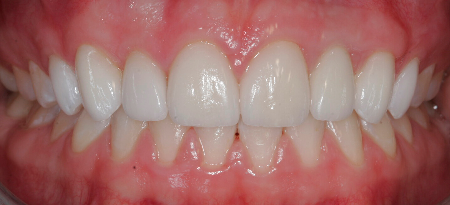
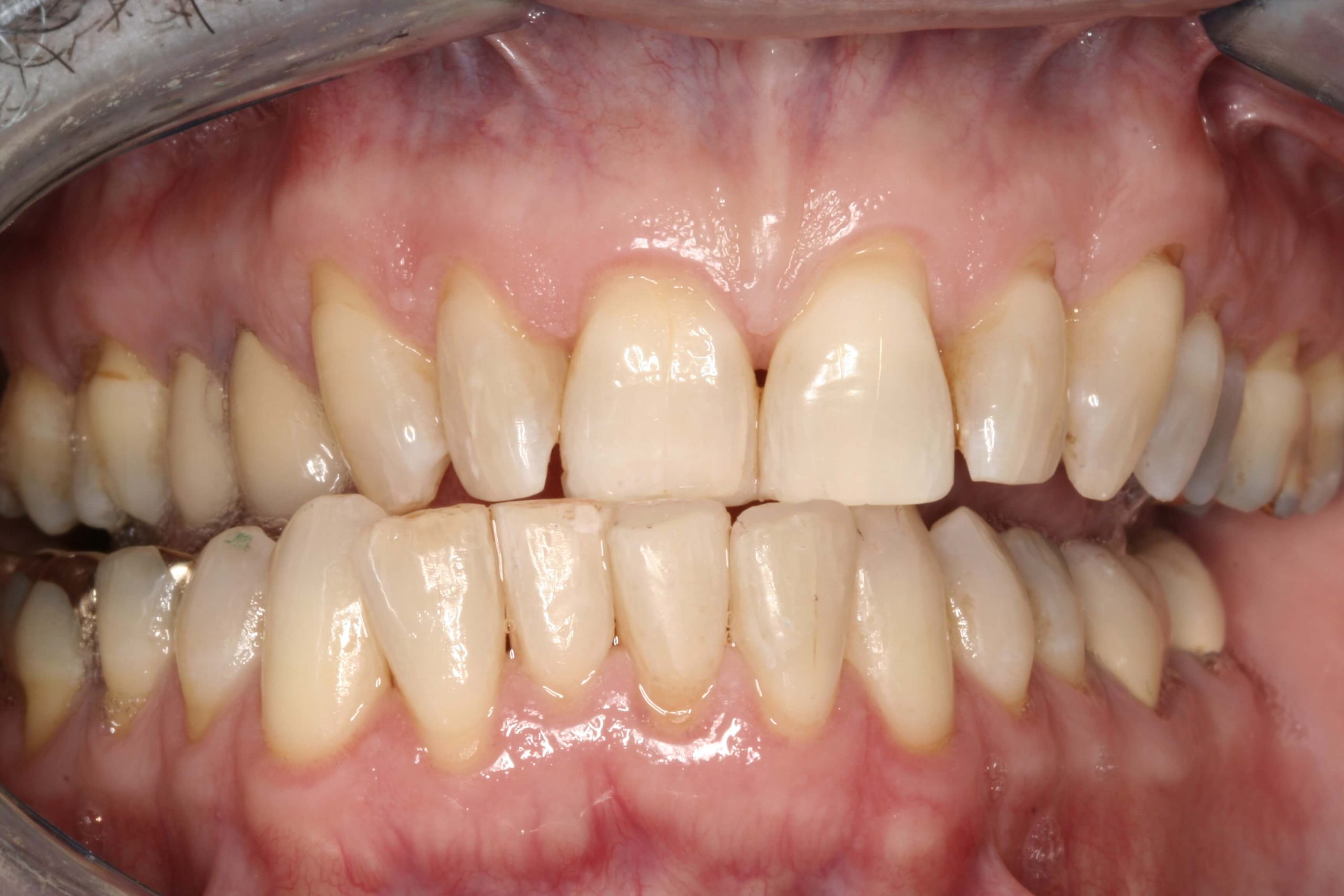
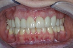
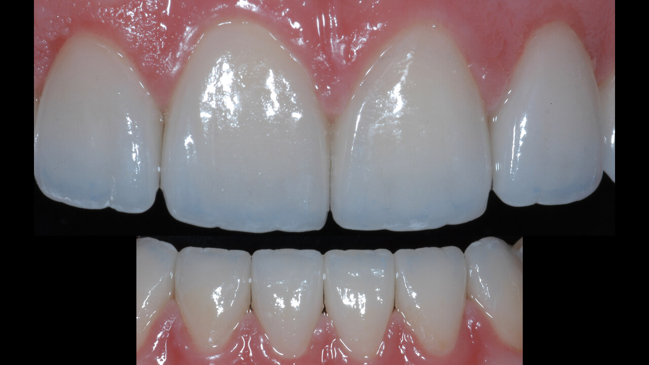
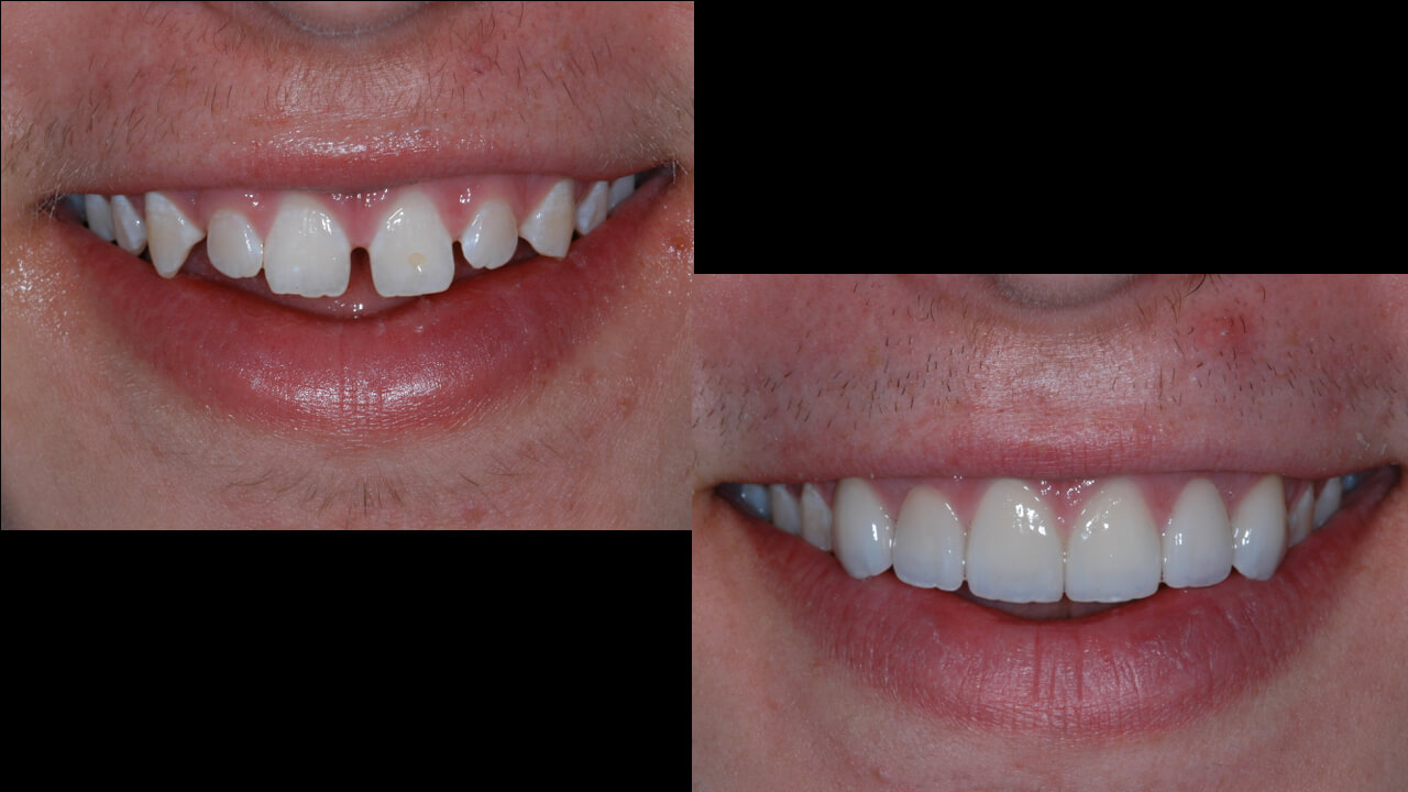
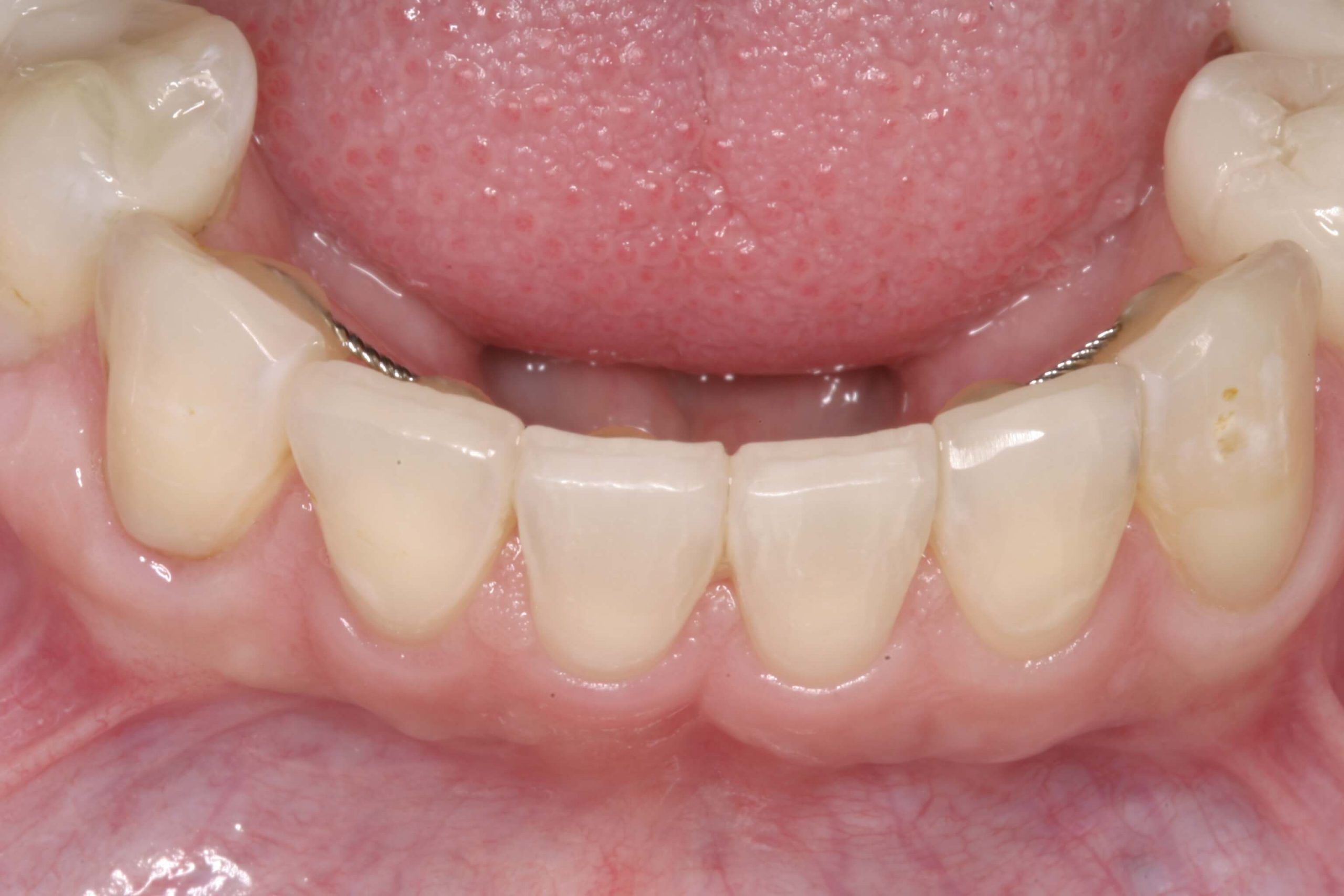
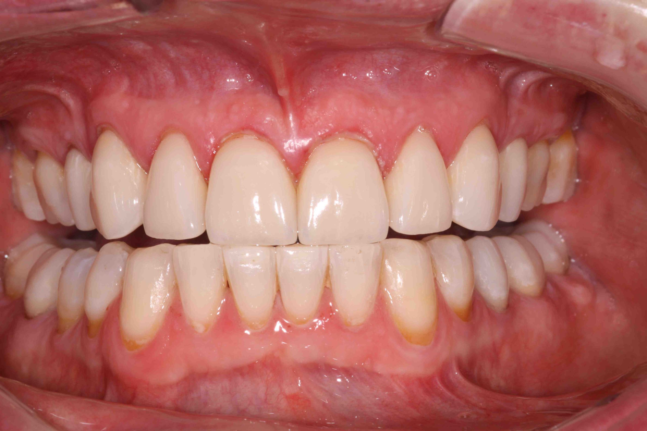
The S Curve
An important tenant of dental aesthetics is the ‘S’ shaped curve that visually stimulates a sense of beauty. This has to do with the way it moves the eye and creates a flowing movement. The curve is a common aesthetic aspect of teeth and tissue, especially in the tips of every papilla to the zenith point.
We see the S shape as a result of the emergence profile or the angle of the entrant line of each tooth. Contour also plays a role in this specific part of the smile’s appearance. All in all, it’s important to pay attention to this aesthetic nuance in your work.
What do you think is the most important consideration for aesthetics in dentistry?


Related Course
E4: Posterior Reconstruction and Completing the Comprehensive Treatment Sequence
DATE: November 7 2024 @ 8:00 am - November 11 2024 @ 2:30 pmLocation: The Pankey Institute
CE HOURS: 44
Dentist Tuition: $ 7300
Single Occupancy with Ensuite Private Bath (per night): $ 290
THIS COURSE IS SOLD OUT The purpose of this course is to help you develop mastery with complex cases involving advanced restorative procedures, precise sequencing and interdisciplinary coordination. Building on…
Learn More>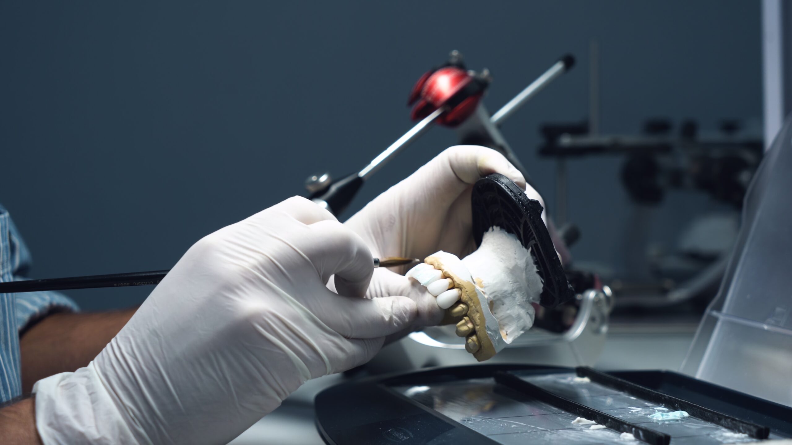
About Author