Using X-Floss for Dental Implant Care
By Lee Ann Brady, DMD
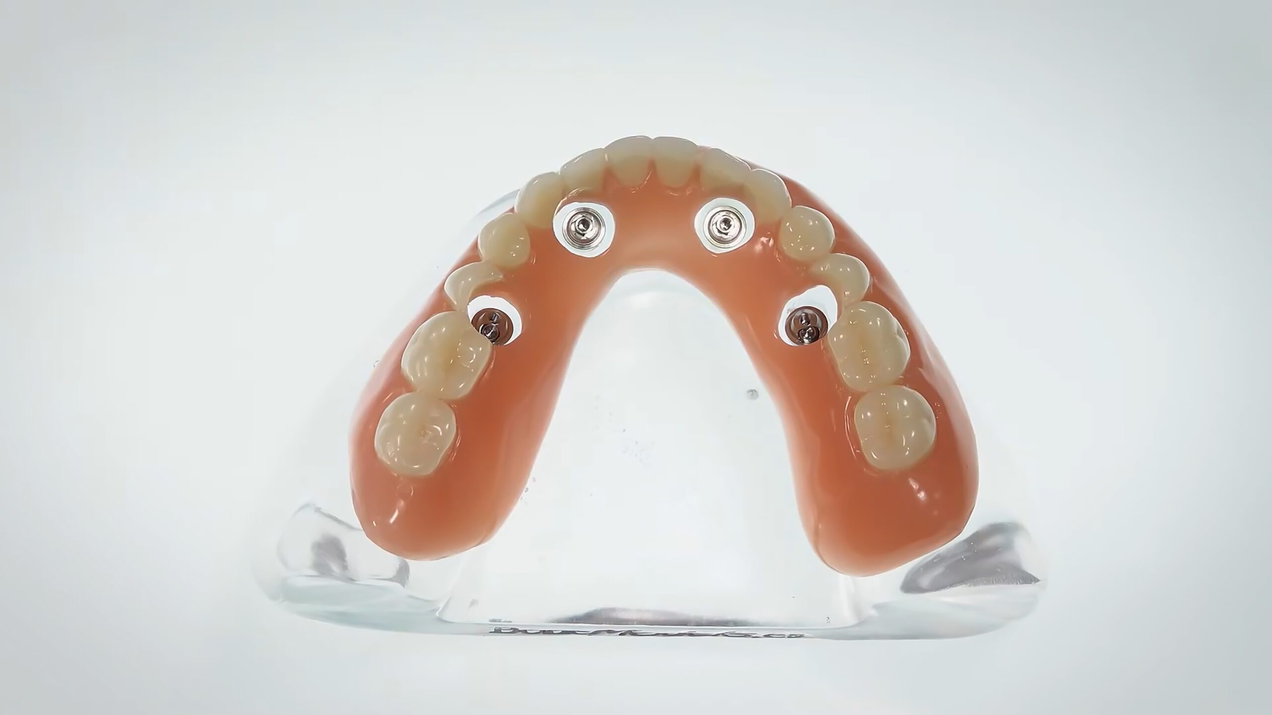
Cleaning the larger gingival embrasures around a posterior dental implant can be a challenge for patients. In my practice, posterior implant patients are some of the individuals we give X-Floss samples to try at home.
X-Floss is a dental floss made by iDontix® that is designed to make flossing easier for individuals with bridges, braces, implants, or larger-than-normal gingival embrasures. It resembles yarn, has a thick texture, and has a hard end, making it easy to push under orthodontic wires, bridges, or in embrasure spaces. It effectively cleans larger spaces while remaining gentle on the gums. The soft material minimizes the risk of injury during flossing, even in subgingival areas, and it is conveniently available on Amazon and in drugstores.
There are two varieties. Green X-Floss from is too thick for some spaces. Blue X-Floss Lite is less thick and just right for some spaces. You and your hygienist may want to give samples of both to your patients to try. Some of your patients are likely to more effectively and consistently floss once they are using this type of floss.


Related Course

Surgically Facilitated Orthodontic Therapy
DATE: October 10 2024 @ 8:00 pm - October 10 2024 @ 9:00 pmLocation: Online
CE HOURS: 1
Date: October 10, 2024 Time: 8 – 9 pm ET Speaker: George Mandelaris, DDS, MS COURSE DESCRIPION Patients seeking ideal esthetics may require a more sophisticated diagnosis and treatment plan…
Learn More>Related Article

About Author
Dr. Lee Ann Brady is passionate about dentistry, her family and making a difference. She is a general dentist and owns a practice in Glendale, AZ limited to restorative dentistry. Lee’s passion for dental education began as a CE junkie herself, pursuing lots of advanced continuing education focused on Restorative and Occlusion. In 2005, she became a full time resident faculty member for The Pankey Institute, and was promoted to Clinical Director in 2006. Lee joined Spear Education as Executive VP of Education in the fall of 2008 to teach and coordinate the educational curriculum. In June of 2011, she left Spear Education, founded leeannbrady.com and joined the dental practice she now owns as an associate. Today, she teaches at dental meetings and study clubs both nationally and internationally, continues to write for dental journals and her website, sits on the editorial board of the Journal of Cosmetic Dentistry, Inside Dentistry and DentalTown Magazines and is the Director of Education for The Pankey Institute.