Digital Splints Today: Part 1

February 28, 2018 Stephen Malone DMD

The new challenge facing us in dentistry is how to incorporate technology into our daily practice. Digital splints specifically are a subject I have been working on for about a year. 

We have had the technology available to mill a splint out of acrylic for a few years now. However, we have not had a good protocol that meets all our needs. 

Digital Splints: Challenges

Some of the problems we face are as follows:

1) Lack of digital articulators that make all of the movements we are able to with semi adjustable articulators, such as crossover transitions. 

2) Absence of centric relation record mountings in software on a computer.

3) No rotational path insertion we can achieve from relines in the mouth. 

4) Few materials that are as good or better than we have now.

I believe we are well on our way to solving these issues. The biggest problem I see is something Dr. Pankey was dealing with many years ago. He talked about how the majority of dentists are indifferent to good comprehensive care dentistry. Therefore, most of the manufacturers of our dental equipment and software are catering to a majority that does not share our own clinical demands. 

These companies give me answers like, “That sounds great doc but who will I be able to sell that to?” I think we have to find workarounds for now that will encourage development in these technologies. Keep in mind, all of the workarounds I will explain are in line with what we teach at the Pankey Institute. 

Digital Splints: Opportunities

We also need systems we can duplicate and teach without compromising the quality of care or experience for patients. I believe there is great potential for higher quality materials and great fitting splints without relines. These two potentials alone can create more value and better experiences for patients.

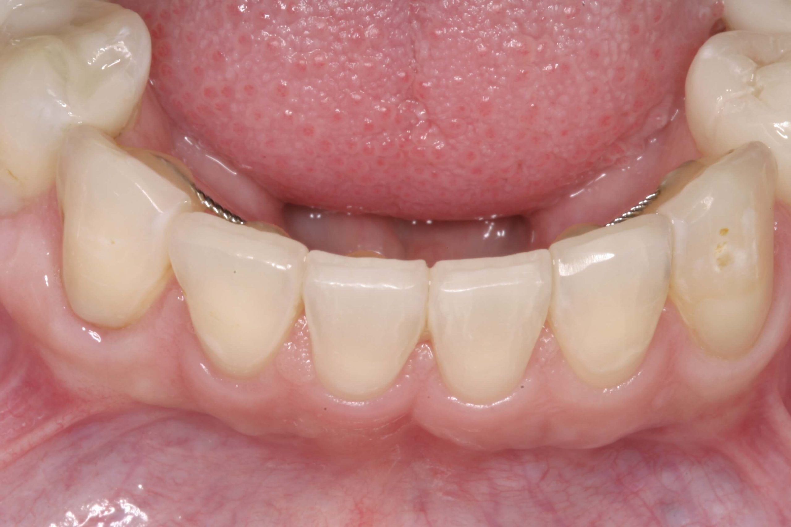
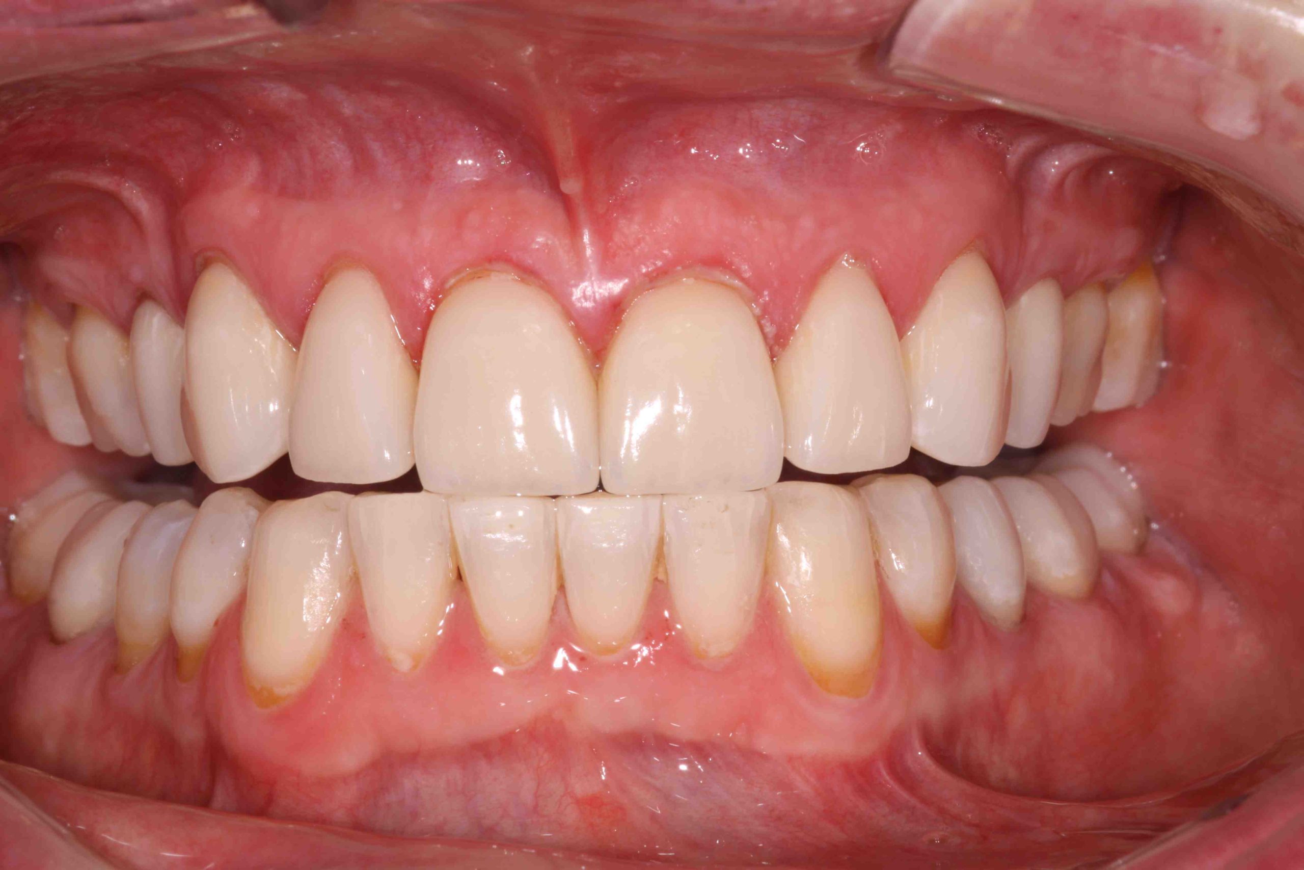
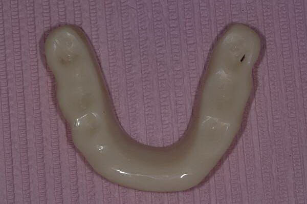
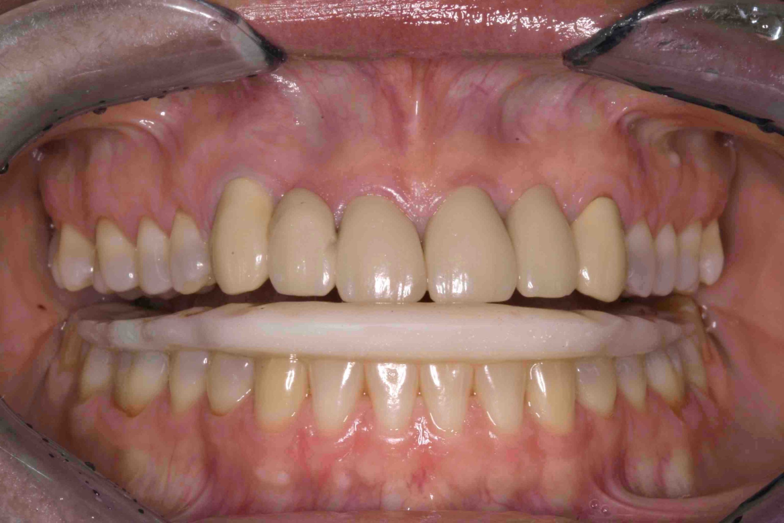
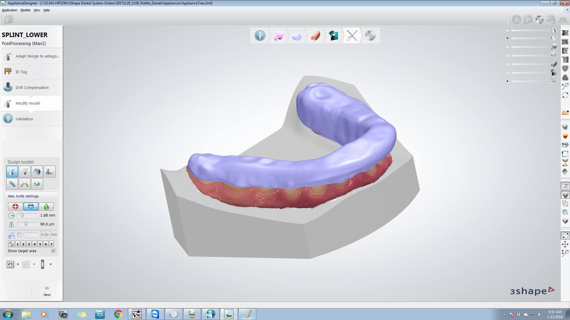
Today I have a protocol that is some digital and some analog. I intraoral scan our impressions with the TRIOS scanner. I believe most of the scanners on the market today work very well and produce very accurate files that can be printed into models. I also use the TRIOS because it communicates very well with the 3SHAPE units most labs use. 

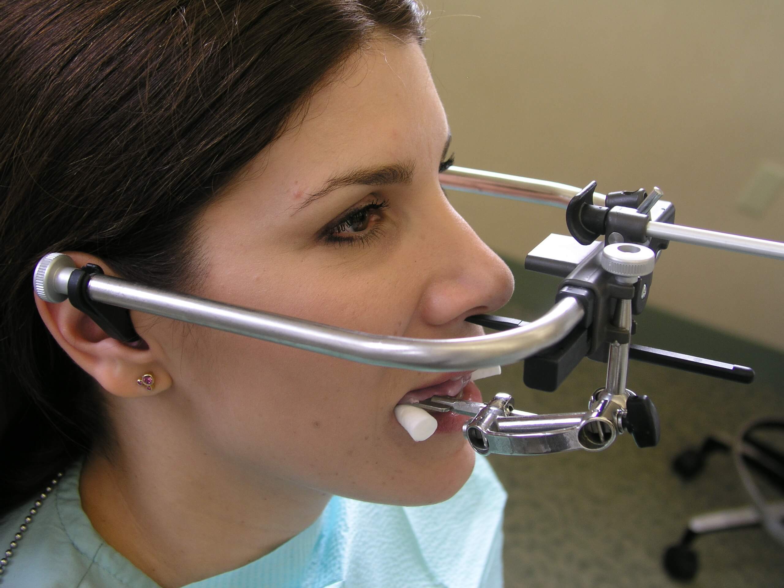
Now that I have files and models I have to mount them. This is our first problem to solve. I still use an analog facebow or facial analyzer. I mount these models on an articulator like the Denar Mark 330 because this is an articulator model programmed into the 3SHAPE software. 

To be continued…

Related Course

E1: Aesthetic & Functional Treatment Planning at the Chicago Midwinter Dental Meeting

DATE: February 20 2025 @ 7:00 am - February 22 2025 @ 8:00 pm

Location: Chicago Midwinter Meeting

CE HOURS:

Transform your experience of practicing dentistry, increase predictability, profitability and fulfillment. The Essentials Series is the Key, and Aesthetic and Functional Treatment Planning is where your journey begins.  Following a system of…

Learn More>

About Author

User Image
Stephen Malone DMD

Dr. Stephen Malone received his Doctorate of Dental Medicine Degree from the University of Louisville in 1994 and has practiced dentistry in Knoxville for nearly 20 years. He participates in multiple dental study clubs and professional organizations, where he has taken a leadership role. Among the continuing education programs he has attended, The Pankey Institute for Advanced Dental Education is noteworthy. He was the youngest dentist to earn the status of Pankey Scholar at this world-renowned post-doctoral educational institution, and he is now a member of its Visiting Faculty.

FIND A PANKEY DENTIST OR TECHNICIAN

I AM A
I AM INTERESTED IN

VIEW COURSE CALENDAR

Leading and Trailing Edges: Part 2

February 26, 2018 Richard Green DDS MBA

How do you ensure aspects like pitch, bevel, crossover, and trailing edges are taken into account? Here, Dr. Green continues his commentary on this valuable aspect of dentistry.

A Flaw of Design: Why Leading & Trailing Edges Matter

While I was helping in the Pankey Scholar Program, the participants would video their patient during the post-case conversation. They would record the movements of the patient in protrusive, left, right radial lateral into crossover, and lateral protrusive.

As I was observing patients in the videos, I would occasionally notice a hesitation once they got to the tips of the cuspid. Sometimes there would also be a quiver of the jaw or muscle twitch. I would usually review the video later with the participant and look at the finished case on the articulator with them. We would talk about how they could put a ‘landing facet’ on cuspids too since they are anterior teeth.

They were beautiful porcelain cases, so I would show them on another set of models how easy it was to do. I told them they would know it was right when the patient’s eyes smiled and the hesitation and muscle twitching went away. Another benefit of a facet on a cuspid is that it is gentler on opposing incisal edges of centrals and laterals in all mandibular movements.

Talking About Edges

One way to talk about the leading and trailing edges: on upper anterior teeth, including cuspids, the leading bevel is at the lingual-incisal junction. The bevel develops naturally on natural teeth. With restorations, the dentist creates it.

The leading edge of the lower incisors, including cuspids, is on the facial incisal junction. It is created in natural teeth with normal function. When restorative material is used, it must be managed by the dentist.

The trailing edges and bevels (labial-incisal edge of upper anteriors plus cuspid and lingual-incisal edges of lower anteriors, including cuspids) are always shaping with function. This can lead to micro-fracturing or major sheering of enamel vertically. Therefore, the trailing edge bevel must always be managed by the dentist with intention. The goal should be preventive with natural tooth or any restorative material.

Once a natural tooth has been worn to the point of losing incisal embrasure, the medial and lingual marginal ridge convex Shaw on the lower can act like a chisel against the labial incisal edge of the upper. This is seen often in a crossover position.

Edges, bevels, and pitch may not be simple, but awareness comes in the doing and observing!

Related Course

E1: Aesthetic & Functional Treatment Planning

DATE: July 17 2025 @ 8:00 am - July 20 2025 @ 2:30 pm

Location: The Pankey Institute

CE HOURS: 39

Dentist Tuition: $ 6800

Single Occupancy with Ensuite Private Bath (Per Night): $ 345

Transform your experience of practicing dentistry, increase predictability, profitability and fulfillment. The Essentials Series is the Key, and Aesthetic and Functional Treatment Planning is where your journey begins.  Following a system of…

Learn More>

About Author

User Image
Richard Green DDS MBA

Rich Green, D.D.S., M.B.A. is the founder and Director Emeritus of The Pankey Institute Business Systems Development program. He retired from The Pankey Institute in 2004. He has created Evergreen Consulting Group, Inc. www.evergreenconsultinggroup.com, to continue his work encouraging and assisting dentists in making the personal choices that will shape their practices according to their personal vision of success to achieve their preferred future in dentistry. Rich Green received his dental degree from Northwestern University in 1966. He was a early colleague and student of Bob Barkley in Illinois. He had frequent contact with Bob Barkley because of his interest in the behavioral aspects of dentistry. Rich Green has been associated with The Pankey Institute since its inception, first as a student, then as a Visiting Faculty member beginning in 1974, and finally joining the Institute full time in 1994. While maintaining his practice in Hinsdale, IL, Rich Green became involved in the management aspects of dentistry and, in 1981, joined Selection Research Corporation (an affiliate of The Gallup Organization) as an associate. This relationship and his interest in management led to his graduation in 1992 with a Masters in Business Administration from the Keller Graduate School in Chicago.

FIND A PANKEY DENTIST OR TECHNICIAN

I AM A
I AM INTERESTED IN

VIEW COURSE CALENDAR

Leading and Trailing Edges: Part 1

February 22, 2018 Richard Green DDS MBA

When Dr. Richard Green read Dr. Lee Ann Brady’s blogs on pitch and bevels, he decided to jump in with a thoughtful response. Read on for more discussion of this fascinating topic.

Crossover and Movement: Consider the Edges

I loved Dr. Brady’s article concerning edges (pitch and both bevels) and the conversation about natural teeth, composition, and porcelain. I was reminded of some of my learning with and from Henry Tanner while refining my occlusion in the mid-70s. It worked for me no matter what material and bite splints were used.

Henry was the first to introduce me to ‘crossover.’ At the time, one of the anterior teeth you did not talk about were the cuspids. They too have important facets (pitch and two bevels) that need to match cusp tip to cusp tip, regardless of the material.

When moving into crossover and the cuspid tips touch, if the pitch facet does not match or is pointed, sloped, or rounded, you often see the masseter or temporalysis muscle twitch. This occurs as the patient hesitates in their movement. That smooth transition back to the incisal edges of the centrals and laterals is important.

I also realized during my career that certain patients (teens, golfers, baseball players) would often stabilize their head while their teeth were cuspid tip to tip or in a crossover position at the point of their impact with the ball.

Improvements can be accomplished by simply taking the flat portion of a ½ J (wheel fine diamond) and creating matching facets on upper and lower cuspids. Polish them and both the leading and trailing bevels so that the movement becomes smooth. If the patient wants to stop cusp tip to cusp tip on the upper and lower cuspid, there is a stable stop and the muscles are comfortable.

To be continued…

Related Course

E1: Aesthetic & Functional Treatment Planning

DATE: October 3 2024 @ 8:00 am - October 6 2024 @ 2:30 pm

Location: The Pankey Institute

CE HOURS: 39

Dentist Tuition: $ 6500

Single Occupancy Room with Ensuite Bath (Per Night): $ 290

Transform your experience of practicing dentistry, increase predictability, profitability and fulfillment. The Essentials Series is the Key, and Aesthetic and Functional Treatment Planning is where your journey begins.  Following a system of…

Learn More>

About Author

User Image
Richard Green DDS MBA

Rich Green, D.D.S., M.B.A. is the founder and Director Emeritus of The Pankey Institute Business Systems Development program. He retired from The Pankey Institute in 2004. He has created Evergreen Consulting Group, Inc. www.evergreenconsultinggroup.com, to continue his work encouraging and assisting dentists in making the personal choices that will shape their practices according to their personal vision of success to achieve their preferred future in dentistry. Rich Green received his dental degree from Northwestern University in 1966. He was a early colleague and student of Bob Barkley in Illinois. He had frequent contact with Bob Barkley because of his interest in the behavioral aspects of dentistry. Rich Green has been associated with The Pankey Institute since its inception, first as a student, then as a Visiting Faculty member beginning in 1974, and finally joining the Institute full time in 1994. While maintaining his practice in Hinsdale, IL, Rich Green became involved in the management aspects of dentistry and, in 1981, joined Selection Research Corporation (an affiliate of The Gallup Organization) as an associate. This relationship and his interest in management led to his graduation in 1992 with a Masters in Business Administration from the Keller Graduate School in Chicago.

FIND A PANKEY DENTIST OR TECHNICIAN

I AM A
I AM INTERESTED IN

VIEW COURSE CALENDAR

TemporomandibularJoint Exam Refresher

February 13, 2018 Lee Ann Brady DMD

The comprehensive exam sets the stage for the quality of your dentistry. The information you gather is instrumental in guiding your treatment plan, getting to know your patient, and helping you effectively relieve pain or discomfort.

The temporomandibular joint is one of the 4 exam areas that comprise a comprehensive functional exam. Ascertaining where we believe the disc is relative to the condyle and whether or not we detect the presence of inflammation are the goals. We want to understand if the joint is stable, adapted or currently undergoing breakdown.

Refresh Your Joint Exam Technique

A good place to start is with lateral pole location. While the patient is lying back, place three fingers lightly in the lateral pole region. Then have them open and close. As they are opening and closing, locate the lateral poles. Observe and record palpable joint noise sounds and motion. Make sure you are documenting your findings clearly throughout the process.

You should also reference maxillary midline to mandibular midline and record opening and closing deviations from the midline. There is so much that can be learned from this basic exam protocol.

Next, move on to joint auscultation in translation and excursions. Using your stethoscope to listen, you can direct the patient to again open and close without touching, as well as move their jaw excursively. You’ll verify palpable sounds and listen to both rotation and translation…

What do you consider critical elements of a joint and muscle exam? 

Related Course

The Pankey Assistant’s Experience

DATE: June 12 2025 @ 12:00 pm - June 14 2025 @ 7:00 pm

Location: The Pankey Institute

CE HOURS: 17

Regular Tuition: $ 2050

Single Occupancy Room with Ensuite Bath (Per Night): $ 345

This “can’t miss” course will empower Dental Assistants to bring their skills to excellence! During this dynamic hands-on course, led by Pankey clinical team member, Sandra Caicedo, participants will learn…

Learn More>

About Author

User Image
Lee Ann Brady DMD

Dr. Lee Ann Brady is passionate about dentistry, her family and making a difference. She is a general dentist and owns a practice in Glendale, AZ limited to restorative dentistry. Lee’s passion for dental education began as a CE junkie herself, pursuing lots of advanced continuing education focused on Restorative and Occlusion. In 2005, she became a full time resident faculty member for The Pankey Institute, and was promoted to Clinical Director in 2006. Lee joined Spear Education as Executive VP of Education in the fall of 2008 to teach and coordinate the educational curriculum. In June of 2011, she left Spear Education, founded leeannbrady.com and joined the dental practice she now owns as an associate. Today, she teaches at dental meetings and study clubs both nationally and internationally, continues to write for dental journals and her website, sits on the editorial board of the Journal of Cosmetic Dentistry, Inside Dentistry and DentalTown Magazines and is the Director of Education for The Pankey Institute.

FIND A PANKEY DENTIST OR TECHNICIAN

I AM A
I AM INTERESTED IN

VIEW COURSE CALENDAR

Who Captures The Facebow Record?

February 10, 2018 Roger Macias DDS

Do you feel reticent about having someone other than you use the facebow? 

A Spatial Reference Point Story

Recently over the holidays as I was “channel surfing” I came across the movie Apollo 13. This is one of those movies that no matter how many times I have watched it, I just have to stop and watch it one more time. Every time I do, I can’t help but get misty-eyed when it gets to the part when the crew make it back to Earth safely (SPOILER ALERT … But you probably read this in the history books anyway).

For me, there is one super exciting moment in the film when Astronaut James Lovell (aka Tom Hanks) has to find a reference point to correct their descent back to Earth from space or burn up on re-entry. Since he cannot use his on-board computers, he lines up his spaceship with the Earth in his window.

“Keep the Earth in the window!” A spatial reference point! Too much correction and their spaceship burns up on re-entry. Too little and they skip off the Earth’s atmosphere.

Make Your Facebow Process Simpler

In our dental offices, the facebow is used to give us a spatial reference point for mounting diagnostic or working models of our patient’s dentition. This is done onto an articulator that approximates the realities of our patient. Sure, you might be able to mount casts arbitrarily, but is your accuracy reproducible? The facebow is a simple tool in our armamentarium to make our life easier.

The question remains, “Is this a task that the dentist must perform?” In my office when we create exquisite dental mountings, I delegate this task to my awesome dental assistants.  With a little training they can do this immediately and the procedure only takes a few minutes.

This involvement is a great way for them to demonstrate their knowledge. It paves the way for more opportunities to open conversations about the Dentist’s Care, Skill, and Judgement. They become your chairside cheerleader and highlight your expertise. They will also express how a critical bite registration record or protrusive record performed by the dentist will only enhance the outcome of treatment.  

Information gathered through the use of a facebow makes our dentistry more predictable. It distinguishes you and your team as a highly trained dental practice.

Don’t burn up on re-entry or skip off into space. Glide effortlessly into beautiful predictable dentistry by using your facebow. Keep your Earth in the window!

Related Course

The Pankey Administrative Team: Inspiring Front Office Excellence

DATE: February 6 2025 @ 8:00 am - February 7 2025 @ 2:00 pm

Location: The Pankey Institute

CE HOURS: 16

Regular Tuition: $ 2150

Single Occupancy Room with Ensuite Bath (Per Night): $ 345

The Pankey Administrative Team: Inspiring front office excellence Front office systems for a Pankey-trained dentist hold very specific differences compared to a  typical dental office.  Learning how to differentiate your…

Learn More>

About Author

User Image
Roger Macias DDS

Dr. Macias obtained his dental education at the University of Texas Health Science Center Dental School at San Antonio and graduated in 1983. While establishing his private practice, Dr. Macias was an assistant professor in the Department of General Practice at the UT Dental School from 1983 until 1989. He is the team dentist for the San Antonio Rampage, the WNBA San Antonio Silver Stars, the San Antonio Missions Baseball Club as well as numerous college universities and high schools in the south Texas area. Dr. Macias is active in numerous dental study clubs and is currently a faculty professor at the world renowned L.D. Pankey Institute for Advanced Dental Education in Key Biscayne, Florida. Among Dr. Macias’s many accolades and awards, he has received his Fellowship in the American and the International College of Dentistry as well as the Pierre Fauchard Academy.

FIND A PANKEY DENTIST OR TECHNICIAN

I AM A
I AM INTERESTED IN

VIEW COURSE CALENDAR

Case Report: Ceramic Veneers & Invisalign Part Two

January 3, 2018 Mike Crete DDS

This is part 2 in a series where Dr. Mike Crete describes an conservative esthetic case where he combined Invisalign and veneers.  Part one looks at helping the patient become aware of the possibilities. Read on for the conclusion.

A Smile Dialogue

… All of this conversation took place in about 5 minutes at the end of the patient’s initial hygiene examination. I then invited him to return for a ‘complimentary smile analysis’ appointment where I would take a series of digital photos and then sit down with him and talk about what we could do to improve his smile.   

I find I can build trust and credibility with a new patient by offering to see them for this complimentary appointment. It only takes about 20 minutes and I typically ‘convert’ the patient to a records appointment (comprehensive exam, X-rays, and mounted study models).

The records appointment was scheduled. More co-discovery revealed how significant Drew’s self-esteem was impacted by his smile and his ‘baby face and baby teeth.’ The records appointment was followed by a diagnostic wax up (or a “3D Design” as I like to call it when talking to patients).

Then a consultation was done to review treatment options. This was a formal case presentation using Powerpoint, photos, and mounted models. Drew’s mother sat in on the consultation appointment.

By having accurately mounted study models on a semi-adjustable articulator, I was able to determine I could give Drew an ideal occlusion AND a pleasing smile. This would involve some minor tooth movement using Invisalign for 6 months and then restoring his upper and lower anterior teeth with conservative porcelain veneers. His posterior teeth were equilibrated during the restorative process. Also, an upper bite guard was fabricated for nighttime wear and added protection of the restorations.  

Drew graduated from college approximately 18 months after I first met him. He completed an internship during his final semester and then was hired by the Fortune 500 company immediately following graduation. He recently got married and said to me, “After I had my teeth done everything in my life started to fall into place. I graduated, got a great job, and met the love of my life. Thanks, doc.”  

Changing a smile and changing a life. It’s being able to impact the lives of others in this way that makes it so rewarding to practice dentistry!  

Related Course

E1: Aesthetic & Functional Treatment Planning

DATE: August 21 2025 @ 8:00 am - August 24 2025 @ 2:30 pm

Location: The Pankey Institute

CE HOURS: 39

Dentist Tuition: $ 6800

Single Occupancy with Ensuite Private Bath (Per Night): $ 345

Transform your experience of practicing dentistry, increase predictability, profitability and fulfillment. The Essentials Series is the Key, and Aesthetic and Functional Treatment Planning is where your journey begins.  Following a system of…

Learn More>

About Author

User Image
Mike Crete DDS

Dr. Mike Crete lives and practices in Grand Rapids, MI. He graduated from the University of Michigan dental school over 30 years ago. He has always been an avid learner and dedicated to advanced continuing education., After completing the entire curriculum at The Pankey Institute, Mike returned to join the visiting faculty. Mike is an active member of the Pankey Board of Directors, teaches in essentials one and runs two local Pankey Learning Groups in Grand Rapids.

FIND A PANKEY DENTIST OR TECHNICIAN

I AM A
I AM INTERESTED IN

VIEW COURSE CALENDAR

Esthetics & Function: Incisal Edge Bevels

December 10, 2017 Lee Ann Brady DMD

There are three critical components to the incisal edge anatomy of anterior teeth. Understanding the function and esthetics of the pitch and two bevels is essential to creating an ideal patient result.

How can a clinician re-create the full anatomic form of the tooth in ceramics and composite? 

In my last blog on this topic, I discussed the dimensions, characterization, esthetics, restorative approach and challenge of mimicking ‘pitch‘ esthetics. Now, I’ll delve into mastering the bevels to create superior restorative results. Combining an esthetic pitch with functional bevels will ensure a smooth composite or ceramic outcome.

Components of Incisal Edge Anatomy Function and Esthetics: Bevels

The two bevels can be found on alternately the labial and the lingual of the transition zone between the pitch and these surfaces. They are often called the leading edge and the trailing edge.

Bevels

Dimensions: The bevels on both sides have a variable width. They can be between less than a millimeter to multiple millimeters long.

Characterization: The bevels lengthen in patients who grind their teeth in an excursive pathway pattern. Patients who parafunction edge to edge might eliminate the bevel. This makes it easier to shear enamel off on the labial or lingual side of the tooth. It also could result in chipping the edge enamel.

Function: The bevel is a transition zone to create smooth functional movement passing from excursive movements onto the pitch. Intercuspal stops on lower incisors are often on or gingival to the bevel.

Whether you are finalizing an equilibration, the occlusion on composites, or ceramics, perfecting anterior guidance is all about both pitch and bevel surfaces. These critical components are a great example of marrying form and function in your technique.

What is your restorative approach for recreating incisal edge anatomy? We’d love to hear from you in the comments! 

Related Course

E1: Aesthetic & Functional Treatment Planning

DATE: June 20 2024 @ 8:00 am - June 23 2024 @ 2:30 pm

Location: The Pankey Institute

CE HOURS: 39

Dentist Tuition: $ 6500

Single Occupancy Room with Ensuite Bath (Per Night): $ 290

THIS COURSE IS SOLD OUT Transform your experience of practicing dentistry, increase predictability, profitability and fulfillment. The Essentials Series is the Key, and Aesthetic and Functional Treatment Planning is where your journey…

Learn More>

About Author

User Image
Lee Ann Brady DMD

Dr. Lee Ann Brady is passionate about dentistry, her family and making a difference. She is a general dentist and owns a practice in Glendale, AZ limited to restorative dentistry. Lee’s passion for dental education began as a CE junkie herself, pursuing lots of advanced continuing education focused on Restorative and Occlusion. In 2005, she became a full time resident faculty member for The Pankey Institute, and was promoted to Clinical Director in 2006. Lee joined Spear Education as Executive VP of Education in the fall of 2008 to teach and coordinate the educational curriculum. In June of 2011, she left Spear Education, founded leeannbrady.com and joined the dental practice she now owns as an associate. Today, she teaches at dental meetings and study clubs both nationally and internationally, continues to write for dental journals and her website, sits on the editorial board of the Journal of Cosmetic Dentistry, Inside Dentistry and DentalTown Magazines and is the Director of Education for The Pankey Institute.

FIND A PANKEY DENTIST OR TECHNICIAN

I AM A
I AM INTERESTED IN

VIEW COURSE CALENDAR

Esthetics & Function: Incisal Edge Pitch

December 8, 2017 Lee Ann Brady DMD

The incisal edge anatomy of anterior teeth is quite complex. This complexity is fundamental to the esthetics of the tooth, as well as the function of incisors. How do we re-create the full anatomic form of the tooth in ceramics and composite? 

Components of Incisal Edge Anatomy Function and Esthetics: Pitch

When the full anatomic form is not precisely recreated, this can lead to esthetic and functional challenges. To successfully mimic this form, the clinician can rely on three components of incisal edges (from a lateral perspective): 1 pitch and 2 bevels.

We can visualize the pitch as the flat top of the incisal edge.

Pitch

Dimensions: Labio-lingual width of at least 1mm that increases from attrition or parafunction in edge to edge position.

Characterization: Pitch is not always parallel to the horizon and its relative position is dependent on the inclination of the incisor. Incisors are inclined just a little bit further to the labial at the incisal edge and the pitch has an upward slant toward the lingual.

Esthetics: The tooth shape and inclination results in an incisal edge esthetic of thinner enamel at the labio-incisal junction. It also creates the highly desirable visual translucence. Leveling the pitch to the horizon can change light reflection which is critical to esthetics of the tooth.

Restorative Approach: Often in ceramics we create a pitch that is level to the horizon and has decreased width of the pitch. This can compromise the esthetics of the translucency, but that can be gained back using stains.

Challenge: The challenge with this shape change in ceramics is that patients often sit in edge to edge position during parafunction. Insufficient pitch width may result in the patient experiencing functional challenges, not finding a comfortable spot to rest and increased parafunctional movement.

I’ll expand on understanding the two bevels in my next incisal edge anatomy blog …

What aspects of incisal edge anatomy do you find most challenging? Let us know in the comments!


Related Course

Functional Esthetic Excellence – Utilizing 100% Digital Workflow

DATE: May 8 2025 @ 8:00 am - May 10 2025 @ 2:00 pm

Location: The Pankey Institute

CE HOURS: 25

Dentist Tuition: $ 3195

Single Occupancy with Ensuite Private Bath (per night): $ 345

Embracing Digital Dentistry This course will introduce each participant to the possibilities of complex case planning utilizing 100% digital workflows. Special emphasis will be placed on understanding how software can…

Learn More>

About Author

User Image
Lee Ann Brady DMD

Dr. Lee Ann Brady is passionate about dentistry, her family and making a difference. She is a general dentist and owns a practice in Glendale, AZ limited to restorative dentistry. Lee’s passion for dental education began as a CE junkie herself, pursuing lots of advanced continuing education focused on Restorative and Occlusion. In 2005, she became a full time resident faculty member for The Pankey Institute, and was promoted to Clinical Director in 2006. Lee joined Spear Education as Executive VP of Education in the fall of 2008 to teach and coordinate the educational curriculum. In June of 2011, she left Spear Education, founded leeannbrady.com and joined the dental practice she now owns as an associate. Today, she teaches at dental meetings and study clubs both nationally and internationally, continues to write for dental journals and her website, sits on the editorial board of the Journal of Cosmetic Dentistry, Inside Dentistry and DentalTown Magazines and is the Director of Education for The Pankey Institute.

FIND A PANKEY DENTIST OR TECHNICIAN

I AM A
I AM INTERESTED IN

VIEW COURSE CALENDAR

Pankey History: The Tanner Appliance

December 7, 2017 Buzz Raymond DDS

Pankey dentists have been instrumental in changing the face of dentistry and launching inspiring innovations that advanced patient care. This post is a continuation of my first blog on the history of Dr. Henry Tanner.

Developing the Tanner Appliance

Dr. Tanner always credited Dr. Ricketts for encouraging him and giving him permission to continue learning what patients were doing with their teeth. They were both using full arch lower appliances, although at that time, Henry simply called his a nightguard. For approximately 15 years, he made that nightguard on a single lower cast and then would adjust it in the mouth.

Some years later at The Pankey Institute, under Dr. John Anderson’s leadership, Dr. Tanner introduced his appliance to Dr. Parker Mahan. Dr. Mahan had a Ph.D. in anatomy, was a full time professor at the University of Florida Dental College, and was a world renowned authority on head and neck pain.  

How Dr. Tanner Changed Dentistry

With the full support and encouragement of Drs. Anderson and Mahan, Henry started teaching about his nightguard using mounted casts. Dr. Anderson initially named it a TANG (Tanner Appliance Night Guard).  

Dr. Tanner always said, “Don’t ever have anything named after you. Whenever people misuse it, then your name is attached.” Over the years, the Tanner appliance has become known as an anatomic appliance. It is extremely individualized, based on the patient’s unique joint, condyle, and tooth anatomy.

The goals are even bilateral centric relation contacts, non-interfering posteriors, and smooth anterior guidance. Dr. Tanner was always curious to see how little could be done to the person’s own unique occlusion to accomplish all those goals. Patient discovery and engagement are keys to Tanner appliance therapy.

Dr. Tanner passed away in 2003. His appliance and his life’s work live on.

Related Course

E2: Occlusal Appliances & Equilibration

DATE: April 6 2025 @ 8:00 am - April 10 2025 @ 2:30 pm

Location: The Pankey Institute

CE HOURS: 44

Dentist Tuition: $ 7400

Single Occupancy with Ensuite Private Bath (per night): $ 345

What if you had one tool that increased comprehensive case acceptance, managed patients with moderate to high functional risk, verified centric relation and treated signs and symptoms of TMD? Appliance…

Learn More>

About Author

User Image
Buzz Raymond DDS

Dr. Buzz serves patients in and around the Minneapolis and St. Paul area at his office in Golden Valley, MN. His goal is to help patients receive the treatment they need for optimal health and aesthetics. Buzz attended the Pankey Institute, and helps teach and mentor other dentists as a member of the faculty. Dr. Buzz’s mentors have included some of the finest dentists in the world. He continues to give back by mentoring and teaching dentists throughout the United States and Canada. In 2013, Dr. Buzz was given the honor of being named a Dental Hero by his peers at the Pankey Institute.

FIND A PANKEY DENTIST OR TECHNICIAN

I AM A
I AM INTERESTED IN

VIEW COURSE CALENDAR

Pankey History: Dr. Henry Tanner

December 6, 2017 Buzz Raymond DDS

Forty five years ago, in the fall of 1972, the first class at The Pankey Institute was held. It’s easy to forget how much things have changed in the world of dental continuing education. Read on for an enlightening reminder of how far dentistry has come in the last few decades … 

Pankey History: Dr. Henry Tanner

Dr. John Anderson and Dr. Loren Miller each had sold their private practices and dedicated their careers to the creation of the Institute. In 1974, Dr. Anderson asked Dr. Henry Tanner to be assistant director of education. Dr. Tanner had been head of fixed prosthodontics at USC School of Dentistry and had made several significant contributions to dentistry.  

Many years earlier, Dr. Tanner had rebuilt the occlusion of Dr. Anderson, who then described Henry as “the finest restorative dentist in the world.” Dr. Henry Tanner is most often associated with the development of the Tanner Appliance.  

Dr. Tanner vividly recalled the first time he made a lower full arch appliance for a woman who was having severe head and face pain. She and her husband went to the hospital emergency room and she was given morphine and Demerol, yet she was not having much relief.

During an emergency visit at his office, Dr. Tanner made an acrylic wafer, placed it directly in her mouth, had her touch it gently with her upper teeth, and asked her to mold it with her tongue. After the acrylic got rubbery, he took it out of her mouth, let it harden, and refined it. She was out of pain the next day.

Within weeks of that experience, Dr. Tanner met a well-recognized orthodontist, Dr. Bob Ricketts, who was taking laminagraphs (sectional x-rays) of his splint patients to monitor condylar position and bony healing in the joint.  

To be continued ...

Related Course

Functional Esthetic Excellence – Utilizing 100% Digital Workflow

DATE: May 8 2025 @ 8:00 am - May 10 2025 @ 2:00 pm

Location: The Pankey Institute

CE HOURS: 25

Dentist Tuition: $ 3195

Single Occupancy with Ensuite Private Bath (per night): $ 345

Embracing Digital Dentistry This course will introduce each participant to the possibilities of complex case planning utilizing 100% digital workflows. Special emphasis will be placed on understanding how software can…

Learn More>

About Author

User Image
Buzz Raymond DDS

Dr. Buzz serves patients in and around the Minneapolis and St. Paul area at his office in Golden Valley, MN. His goal is to help patients receive the treatment they need for optimal health and aesthetics. Buzz attended the Pankey Institute, and helps teach and mentor other dentists as a member of the faculty. Dr. Buzz’s mentors have included some of the finest dentists in the world. He continues to give back by mentoring and teaching dentists throughout the United States and Canada. In 2013, Dr. Buzz was given the honor of being named a Dental Hero by his peers at the Pankey Institute.

FIND A PANKEY DENTIST OR TECHNICIAN

I AM A
I AM INTERESTED IN

VIEW COURSE CALENDAR