Demystifying Occlusion

August 9, 2019 Lee Ann Brady DMD

I’ll admit that for a portion of my professional career I didn’t think twice about occlusion.

Today occlusion is as well integrated into my thought process as caries, perio or restorative considerations. What role does occlusion play in your practice? Is it part of your routine diagnostics? Is it fully integrated with your esthetic and restorative treatment planning? Or do you only wonder about it when a patient breaks something or you are concerned about moving forward with a severe wear case?

Early in my career occlusion would show up as a frustration.

One example is when I would prepare a second molar being very careful about creating adequate occlusal clearance, using both depth-cutting burs and checking the result with bite registration, just to have my assistant come and tell me she didn’t have enough clearance to make the provisional. It was a relief years later in my first CE course with a focus on occlusion to learn this was not rapid super-eruption or a mistake on my part, but muscle release due to removal of a key occlusal contact, and I could predict this before I prepped the tooth.

How about the patient who would come into my office for a hygiene visit or a buccal pit restoration with no joint sounds and call the next day concerned that their jaw had been clicking ever since they left the office the day before? What a relief when I learned these patients had an underlying risk for disc displacement called ligament laxity, and I could diagnose it quickly at an exam appointment.

An everyday occlusal issue I run into is the patient with a limited opening who needs posterior dentistry.

Perhaps, they can open at the beginning but rapidly fatigue and their jaw begins to shake and close as we work. What a gift it is today that I can identify this as a symptom of overuse of the elevator muscles, treat it easily and quickly at a restorative appointment with a deprogrammer, and offer the patient options for relaxing their muscles and allowing them to stay healthy.

The process of demystifying occlusion and having it become an everyday reality for me required committing to a series of hands-on CE programs, being willing to manage my learning, and then taking it back to my patients and beginning to use what I was learning in small steady steps. The benefit has been less frustration, increased confidence with my patients, and an ability to help patients in new and profound ways I didn’t have before.

My appreciation for occlusion didn’t stop with my practice.

It became a passion and is a huge piece of the continuing education I teach with The Pankey Institute to demystify occlusion for others.

Related Course

E1: Aesthetic & Functional Treatment Planning

DATE: December 10 2026 @ 8:00 am - December 13 2026 @ 2:30 pm

Location: The Pankey Institute

CE HOURS: 39

Dentist Tuition: $ 6900

Single Occupancy with Ensuite Private Bath (Per Night): $ 355

Transform your experience of practicing dentistry, increase predictability, profitability and fulfillment. The Essentials Series is the Key, and Aesthetic and Functional Treatment Planning is where your journey begins.  Following a system of…

Learn More>

About Author

User Image
Lee Ann Brady DMD

Dr. Lee Ann Brady is passionate about dentistry, her family and making a difference. She is a general dentist and owns a practice in Glendale, AZ limited to restorative dentistry. Lee’s passion for dental education began as a CE junkie herself, pursuing lots of advanced continuing education focused on Restorative and Occlusion. In 2005, she became a full time resident faculty member for The Pankey Institute, and was promoted to Clinical Director in 2006. Lee joined Spear Education as Executive VP of Education in the fall of 2008 to teach and coordinate the educational curriculum. In June of 2011, she left Spear Education, founded leeannbrady.com and joined the dental practice she now owns as an associate. Today, she teaches at dental meetings and study clubs both nationally and internationally, continues to write for dental journals and her website, sits on the editorial board of the Journal of Cosmetic Dentistry, Inside Dentistry and DentalTown Magazines and is the Director of Education for The Pankey Institute.

FIND A PANKEY DENTIST OR TECHNICIAN

I AM A
I AM INTERESTED IN

VIEW COURSE CALENDAR

When Your Occlusal Clearance Disappears

April 19, 2019 Lee Ann Brady DMD

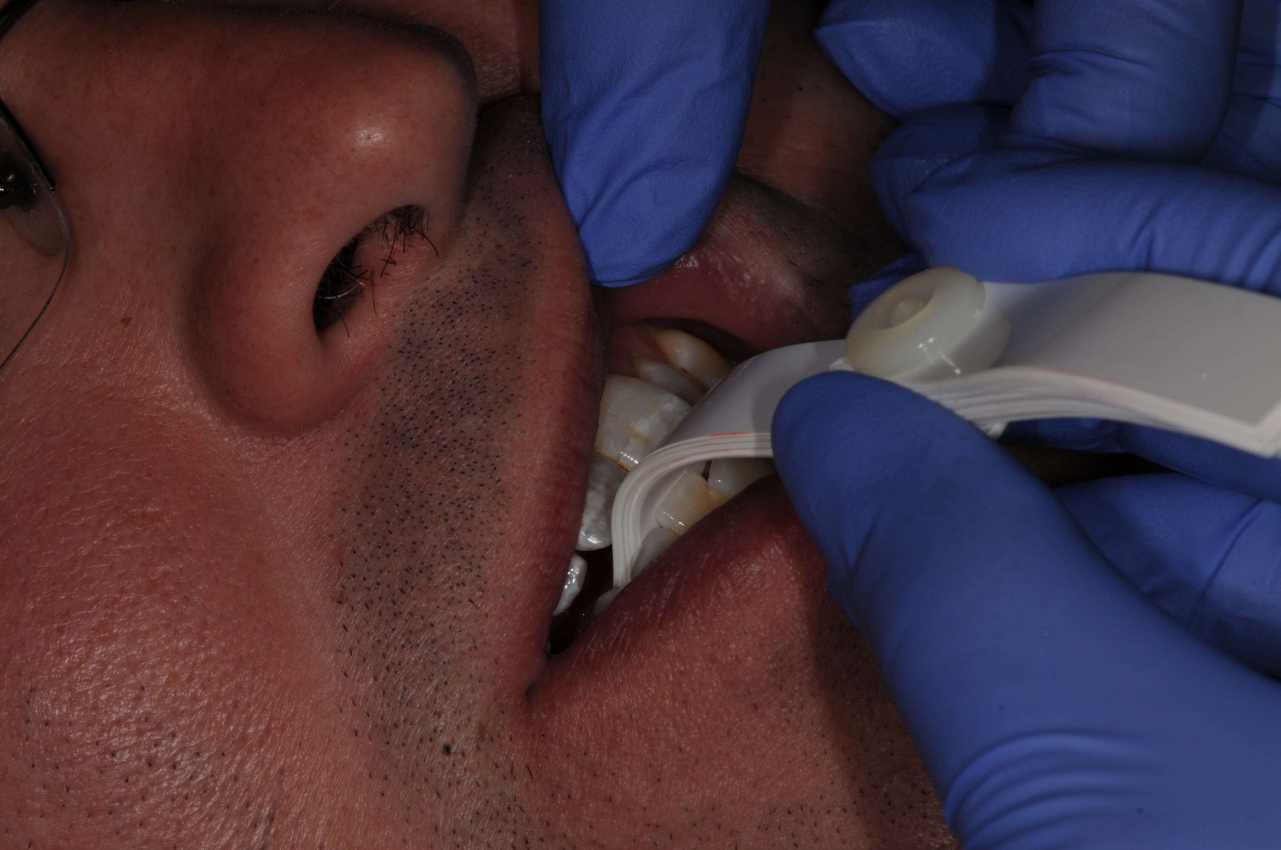
It can be an incredibly frustrating clinical situation, when you have been meticulous about preparing a posterior tooth, (most commonly a molar) for a crown and things aren’t predictable. Using your burs you created depth cuts to ensure adequate occlusal clearance. After the impression you allow your assistant to fabricate the temporary only to have them come get you. Why? Because the temp is thin or perforated on the occlusal. When you go back to check, and have the patient bite, sure enough the opposing tooth is touching your prep.

A common reason that this happens is because we just prepared away the patient’s first point of contact in centric relation. The lateral pterygoid muscle in coordination with the elevator muscles has a learned pattern of firing that bring the mandibular teeth into maximum intercuspal position. This “learned” position is programmed by the patient’s first point of contact when the condyles are seated. For some patients when we remove this contact, and therefore the message that was programming the muscles to locate MIP, they release quickly. When the muscles release and the condyles seat, the occlusion is now totally different than MIP was moments before.

Leaf Gauge
Finding First Point of Contact

One solution that I considered briefly was to no longer work on molars! Alas, not a great business strategy.

Removing this frustration is about understanding which patients are at risk. Identifying risk begins with the exam, whether we are discussing caries or occlusion. There are several key factors that alert me to this potential issue. I start by identifying the patient’s first point of contact and clarifying if it is on the tooth we are about to prepare. If I am going to prepare FPC away, then I look at the magnitude and direction of the patient’s slide, or the difference between this position and MIP. If the difference is small (1-2mm), then even if their condyle does seat the occlusal difference will not cause an issue for clearance. So large slides (3mm or greater) could cost approximately 1mm of clearance on the prepared tooth. Other factors include whether they have a history of occlusal changes or more than one MIP they can find.

Understanding the risk, still leaves us with the question of how to proceed. That is a longer conversation for another post. However, if we proceed as we would before, at least knowing the risk we can explain this to the patient ahead of time, and help them understand how we would manage it if it happens.

Related Course

E3: Restorative Integration of Form & Function

DATE: March 30 2025 @ 8:00 am - April 3 2025 @ 2:30 pm

Location: The Pankey Institute

CE HOURS: 41

Dentist Tuition: $ 7400

Single Occupancy with Ensuite Private Bath (per night): $ 345

THIS COURSE IS SOLD OUT Understanding that “form follows function” is critical for knowing how to blend what looks good with what predictably functions well. E3 is the phase of…

Learn More>

About Author

User Image
Lee Ann Brady DMD

Dr. Lee Ann Brady is passionate about dentistry, her family and making a difference. She is a general dentist and owns a practice in Glendale, AZ limited to restorative dentistry. Lee’s passion for dental education began as a CE junkie herself, pursuing lots of advanced continuing education focused on Restorative and Occlusion. In 2005, she became a full time resident faculty member for The Pankey Institute, and was promoted to Clinical Director in 2006. Lee joined Spear Education as Executive VP of Education in the fall of 2008 to teach and coordinate the educational curriculum. In June of 2011, she left Spear Education, founded leeannbrady.com and joined the dental practice she now owns as an associate. Today, she teaches at dental meetings and study clubs both nationally and internationally, continues to write for dental journals and her website, sits on the editorial board of the Journal of Cosmetic Dentistry, Inside Dentistry and DentalTown Magazines and is the Director of Education for The Pankey Institute.

FIND A PANKEY DENTIST OR TECHNICIAN

I AM A
I AM INTERESTED IN

VIEW COURSE CALENDAR

Do Patients Parafunction In Centric Relation?

April 13, 2019 Lee Ann Brady DMD

Seated Condylar Position

I know even the mention of the words centric relation probably has some readers bristling, as this is a much argued over topic. With that said the research by Lundeen and Gibbs at the University of Florida shows that we do seat our condyles into the fossa during the chewing stroke. This seated condylar position is often used as a reference position to treat patients whether as part of reorganizing their occlusion to alleviate TMD symptoms or for restorative or orthodontic treatments.

Centric Relation & Parafunction

The next question is do people seat their condyles other than during normal function as part of the chewing stroke. I believe the answer is yes. One of the pieces of evidence is the number of patients that I have with wear facets that correspond exactly to their first point of contact with their condyles seated. These same patients do not mark this area with articulating paper in intercuspal position or when following their excursives.

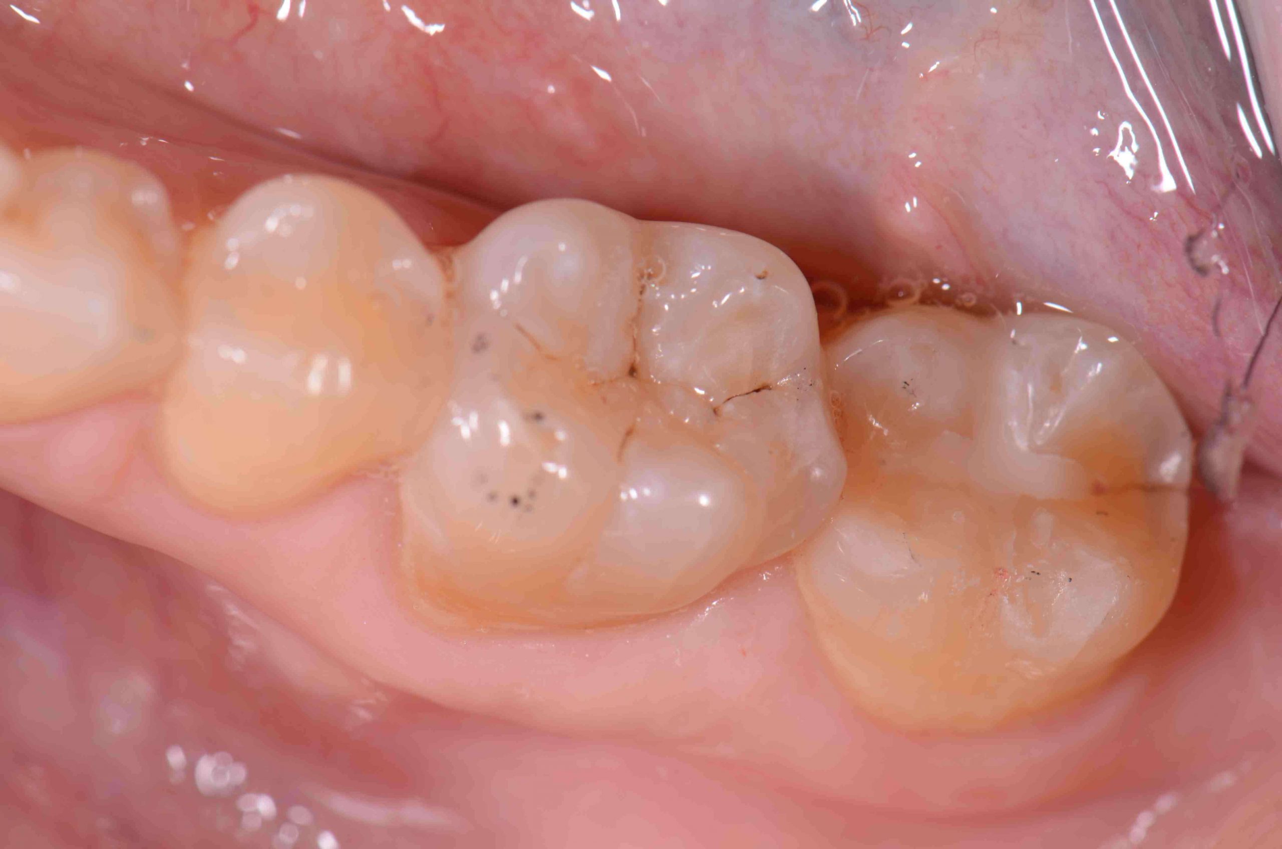
I took the photo with this post in my office. The patient has no other wear facets. #31 has a small, less than .5mm combination sealant/occlusal composite on this tooth. The distal wear facet does not touch in intercuspal position or excursives, but will mark using a leaf gauge to seat the condyles as the first point of contact. This facet marks in both the arc of closure and a power wiggle or abbreviated excursive movement from this first contact. #31 is also split from the gingival margin on the distal over the marginal ridge and right to the margin of the composite. I have seen and restored multiple examples just like this. In my experience when the crown comes back from the lab we will be able to adjust it in without issue, but the patient will report it feels high, or it will become chronically sensitive. The solution will be to either adjust this crown in both intercuspal position and centric relation, or incorporate an equilibration with the restorative care.

My belief is this patient parafunctions in centric relation.

Related Course

E4: Posterior Reconstruction and Completing the Comprehensive Treatment Sequence

DATE: April 23 2026 @ 8:00 am - April 27 2026 @ 2:30 pm

Location: The Pankey Institute

CE HOURS: 44

Dentist Tuition: $ 7500

Single Occupancy with Ensuite Private Bath (per night): $ 355

The purpose of this course is to help you develop mastery with complex cases involving advanced restorative procedures, precise sequencing and interdisciplinary coordination. Building on the learning in Essentials Three…

Learn More>

About Author

User Image
Lee Ann Brady DMD

Dr. Lee Ann Brady is passionate about dentistry, her family and making a difference. She is a general dentist and owns a practice in Glendale, AZ limited to restorative dentistry. Lee’s passion for dental education began as a CE junkie herself, pursuing lots of advanced continuing education focused on Restorative and Occlusion. In 2005, she became a full time resident faculty member for The Pankey Institute, and was promoted to Clinical Director in 2006. Lee joined Spear Education as Executive VP of Education in the fall of 2008 to teach and coordinate the educational curriculum. In June of 2011, she left Spear Education, founded leeannbrady.com and joined the dental practice she now owns as an associate. Today, she teaches at dental meetings and study clubs both nationally and internationally, continues to write for dental journals and her website, sits on the editorial board of the Journal of Cosmetic Dentistry, Inside Dentistry and DentalTown Magazines and is the Director of Education for The Pankey Institute.

FIND A PANKEY DENTIST OR TECHNICIAN

I AM A
I AM INTERESTED IN

VIEW COURSE CALENDAR

Fleximount To Stabilize Lower Model

February 18, 2019 Lee Ann Brady DMD

Learning from one another is one of the top benefits of dental continuing education.

One of the things that I value about continuing dental education is the opportunity to spend time with other dentists.  I always learn something I can bring back to my office. Recently while lecturing at Midwestern Dental School to the faculty, on of the faculty members told me about a new way to stabilize lower models when mounting, and was even kind enough to give me some samples.

Stabilizing a lower model during mounting with centric relation records is critical to the accuracy of the mounting.

Over the years I have tried about every idea possible to optimize mounting the lower model. If the model moves in the bite registration due to pressure during mounting, tipping or shrinkage of the stone it interferes with the accuracy of the mounting. To overcome this I have tried hot glue, compound, rubber bands, hanger wire bent into a V and probably many more.

We realized the Fleximount was incredible the first time we used it. Sold by WhipMix and developed for their articulator systems, I will say I have used it on other systems, and as long as there is a knob on the upper member of the articulator it works fantastic. The Fleximount is trapped inside the stone, so they are disposable. The lower model is held with even pressure directly against the upper( if mounting in MIP) or the bite record, therefore no tipping forces are present as with other stabilizer systems. Because it stays in the stone, you can walk away and let it come to a complete set, instead of having to stand and remove the stabilizer while the mounting stone is still somewhat soft. Both of these features result in a very accurate mounting.

Once the stone is set you simply cut away the rubber band material that is not inside the mounting stone. Now you can finish and groom the lower mounting.

Related Course

TMD & Orofacial Pain: Managing Complex Patients

DATE: January 29 2025 @ 8:00 am - February 2 2025 @ 1:00 pm

Location: The Pankey Institute

CE HOURS: 37

Dentist Tuition: $ 7200

Single Occupancy with Ensuite Private Bath (per night): $ 345

THIS COURSE IS SOLD OUT TMD patients present with a wide range of concerns and symptoms from tension headaches and muscle challenges to significant joint inflammation and breakdown. Accurate thorough…

Learn More>

About Author

User Image
Lee Ann Brady DMD

Dr. Lee Ann Brady is passionate about dentistry, her family and making a difference. She is a general dentist and owns a practice in Glendale, AZ limited to restorative dentistry. Lee’s passion for dental education began as a CE junkie herself, pursuing lots of advanced continuing education focused on Restorative and Occlusion. In 2005, she became a full time resident faculty member for The Pankey Institute, and was promoted to Clinical Director in 2006. Lee joined Spear Education as Executive VP of Education in the fall of 2008 to teach and coordinate the educational curriculum. In June of 2011, she left Spear Education, founded leeannbrady.com and joined the dental practice she now owns as an associate. Today, she teaches at dental meetings and study clubs both nationally and internationally, continues to write for dental journals and her website, sits on the editorial board of the Journal of Cosmetic Dentistry, Inside Dentistry and DentalTown Magazines and is the Director of Education for The Pankey Institute.

FIND A PANKEY DENTIST OR TECHNICIAN

I AM A
I AM INTERESTED IN

VIEW COURSE CALENDAR

Master Wax Centric Relation Bite Record 2

October 15, 2018 Pankey Gram

Now that you have fabricated the platform, the next step is to capture the record with the patient. You will need a heat source and a blue Delar wax Pencil.

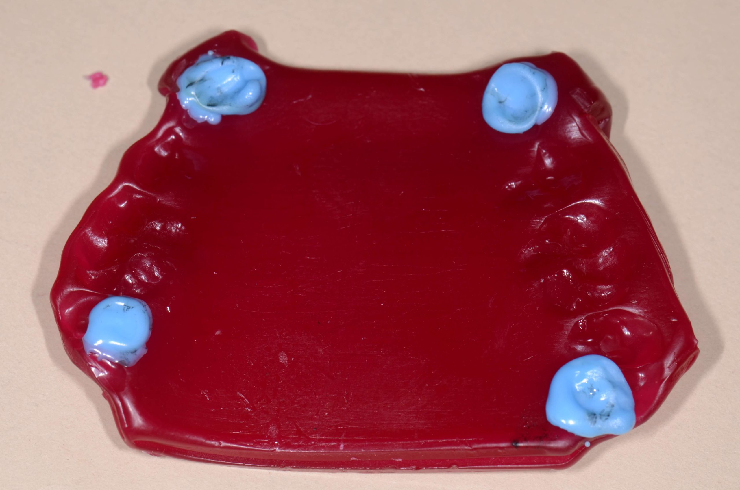
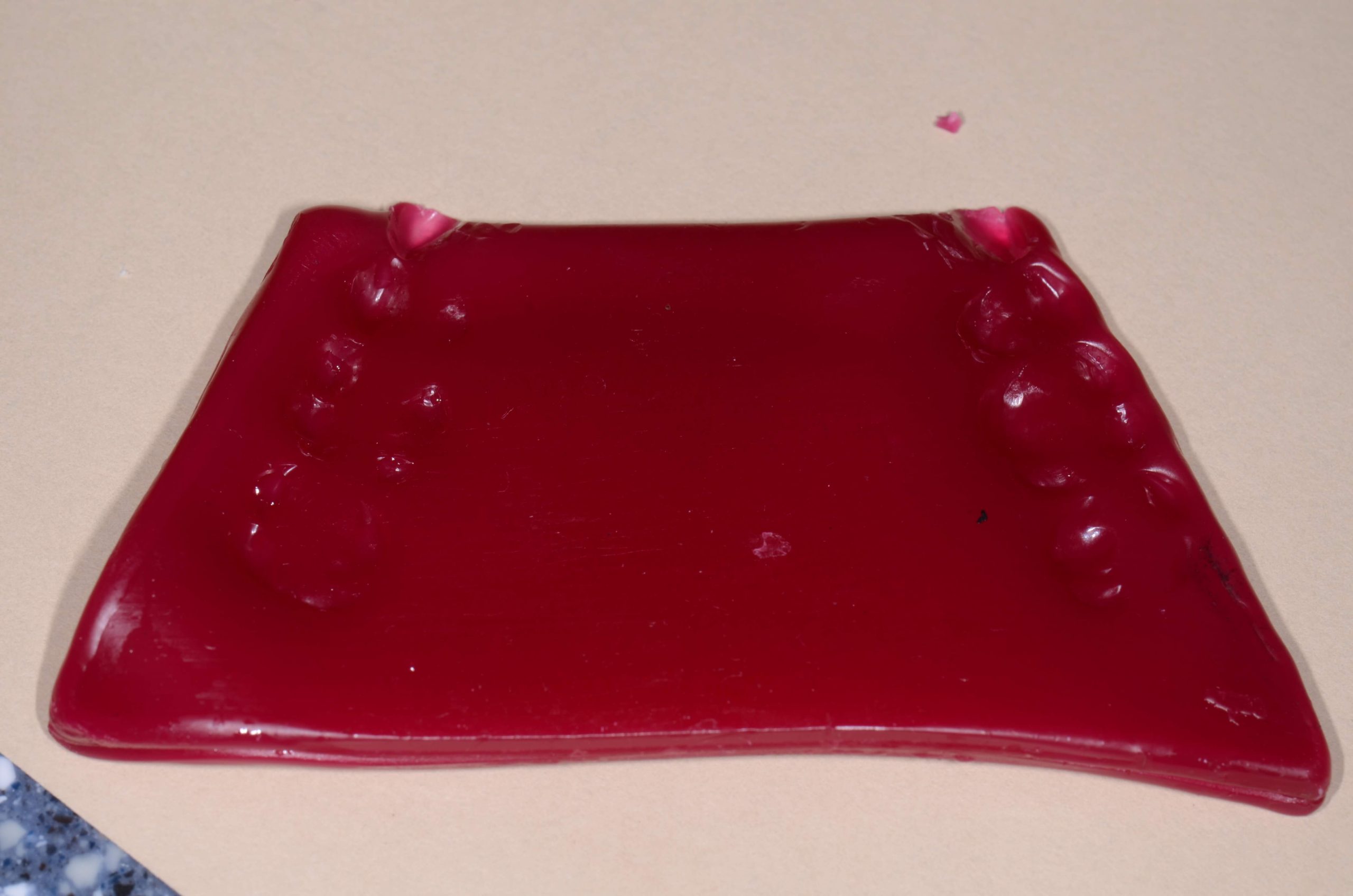
The first step is to heat the sides of the Master Wax Platform so they are tempered. Take the wax to the patient’s mouth and place it over the maxillary teeth. The anterior edge of the wax should be at the embrasure between the canine and the lateral. Bend the corners over the canines to help with retention. Press the wax against the teeth and ask the patient to close gently into the wax. Cool the wax with your air water syringe, have the patient open, and continue to cool the platform before removing from the mouth.

Using blue delar wax created a small bead where the lower canines have left an impression. Reseat the platform over the maxillary teeth and using bimanual guidance bring the lower canine cusp tips up until they just touch the blue wax. Have your assistant cool the wax with air. After removing the platform from the mouth add Delar wax where the second molars have left a cusp imprint. Then return the platform to the mouth and using bimanual guidance arc the patient into the wax so the lower molar cusp touches, then cool with air.

Your record should now be dropped into cool water. A disposable plastic container from the grocery works great. Write the patients name on it with sharpie marker and add it to their lab pan.


Related Course

E2: Occlusal Appliances & Equilibration

DATE: May 3 2025 @ 8:00 am - May 7 2026 @ 2:30 pm

Location: The Pankey Institute

CE HOURS: 44

Dentist Tuition: $ 7500

Single Occupancy with Ensuite Private Bath (per night): $ 355

What if you had one tool that increased comprehensive case acceptance, managed patients with moderate to high functional risk, verified centric relation and treated signs and symptoms of TMD? Appliance…

Learn More>

About Author

User Image
Pankey Gram

FIND A PANKEY DENTIST OR TECHNICIAN

I AM A
I AM INTERESTED IN

VIEW COURSE CALENDAR

Master Wax Centric Relation Bite Record 1

October 12, 2018 Pankey Gram

Analyzing a patient’s occlusion is key to providing optimal care. There are multiple ways to take a centric relation bite record, but one of the classics at Pankey has to be utilizing master wax. Easily mounting models in the laboratory is one of the main advantages of this method.

Although a slightly more challenging technique from a chairside perspective, it can still be accomplished with ease. Here is where to start:

Intro to the Master Wax Centric Relation Bite Record

Begin by gathering your necessary tools and materials. Start with a red master wax that looks just like baseplate wax from a standpoint of the size of the wax sheets, although it is different because it’s both softer and tackier. You will also need a blue wax pencil, scissors to cut the wax, a heat source such as an alcohol torch, and a way to light the heat source.

Take a single sheet of wax and temper it in the middle with the intention of being able to bend it in half. Once it is thoroughly tempered, bend it in half without cracking or breaking the wax. Make sure the bend is fairly crisp. After the wax has cooled a little bit, open it back up and cut it in half. Out of every sheet of wax, you should be able to generate two platforms for wax records.

Once again temper the wax halfway in the middle with the heat source. Tempering refers to heating to flexibility but not dripping. Again, fold the wax in half so you have a double thickness sheet. Then, cut the wax into the shape of the platform …

Look for the second part in this series about our master wax technique coming soon. And did you get a chance to read this Pankey Gram blog on enjoying Miami while you visit Pankey? Let us know what you think!

Related Course

E3: Restorative Integration of Form & Function

DATE: August 16 2026 @ 8:00 am - August 20 2026 @ 2:30 pm

Location: The Pankey Institute

CE HOURS: 41

Dentist Tuition: $ 7500

Single Occupancy with Ensuite Private Bath (per night): $ 355

Understanding that “form follows function” is critical for knowing how to blend what looks good with what predictably functions well. E3 is the phase of your Essentials journey in which…

Learn More>

About Author

User Image
Pankey Gram

FIND A PANKEY DENTIST OR TECHNICIAN

I AM A
I AM INTERESTED IN

VIEW COURSE CALENDAR

Panadent Dento-Facial Analyzer Technique: Level Planes

October 7, 2018 Lee Ann Brady DMD

Function and esthetics are the two primary goals of excellent treatment. Achieving them both simultaneously requires the right tools used with the best skill possible. The  Dento-Facial Analyzer is my go-to for gathering information I can use to improve the outcome of mounting a maxillary model.

In parts 1 and 2 of this series, I introduced the Dento-Facial Analyzer and began the discussion of how to capture records with it. Here, I’ll complete my overview of a solid technique:

Completing the Dento-Facial Analyzer Technique

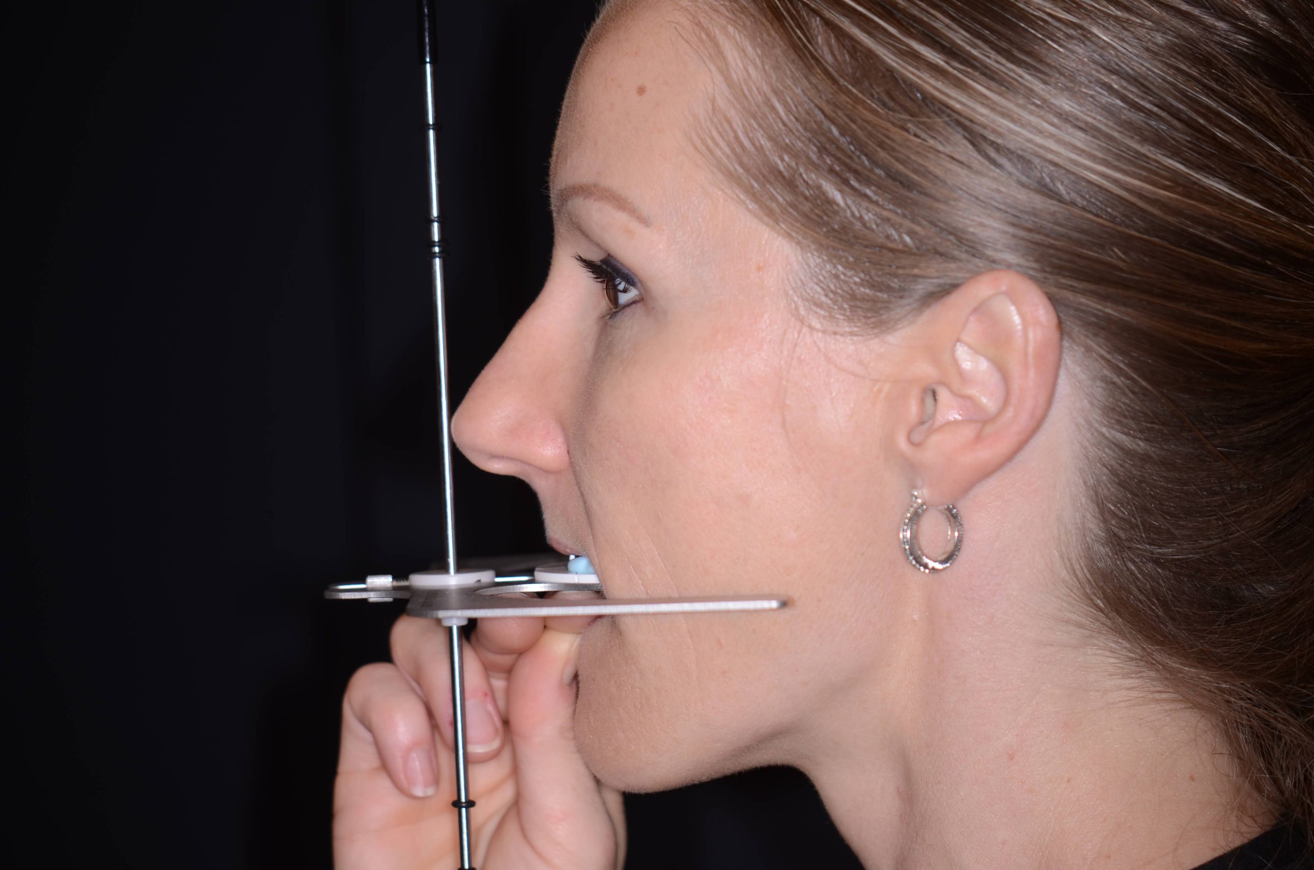
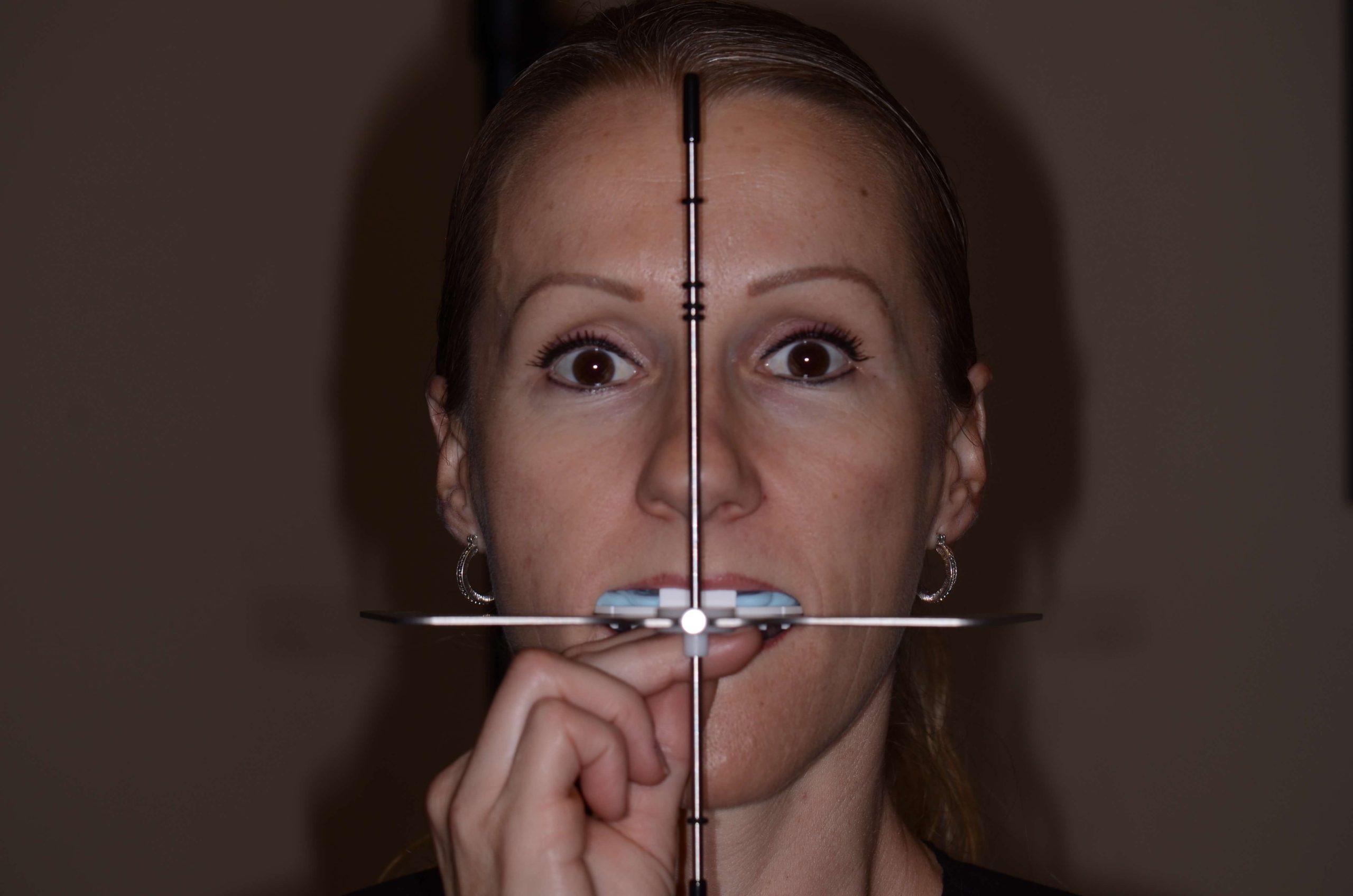
… Ensure the Dento-Facial Analyzer is positioned level to the horizon both when looking straight on at the patient’s face from the anterior section and looking at them from the side. It should be level in both planes of space. Then, allow the bite silicone to set and have the patient hold to verify.

Remember that the main use of the Dento-Facial Analyzer is transferring three significant pieces of information. This is either intended for the laboratory or for when we mount our own models.

The first piece of information is the maxillary relationship – the distance to hinge access – which means it’s very important that the central incisors on the maxilla are seated against the plastic bite plate.

Second, we are transferring information about the occlusal plane and the incisal plane. From an incisal plane perspective, it’s crucial that the plate is level to the horizon as we look straight on at the patient once we have the analyzer in. The vertical rod on the analyzer indicates the center of the face – the facial midline – which can be given by the central philtrum of the upper lip or the center of glabella.

You should also look at how you’ve captured the record from a lateral view. This ensures the occlusal plane – the relationship of the cant from anterior to posterior teeth that exists in the patient’s face – is transferred accurately to the lab or onto the articulator. The side bar of the Dento-Facial Analyzer should be level to the horizon.

Do you use this simple and accurate tool?

For a hands-on demonstration of the Dento-Facial Analyzer from Pankey educators, learn more about our Essentials 1 course.

Related Course

E2: Occlusal Appliances & Equilibration

DATE: May 3 2026 @ 8:00 am - May 7 2026 @ 2:30 pm

Location: The Pankey Institute

CE HOURS: 44

Dentist Tuition: $ 7500

Single Occupancy with Ensuite Private Bath (per night): $ 355

What if you had one tool that increased comprehensive case acceptance, managed patients with moderate to high functional risk, verified centric relation and treated signs and symptoms of TMD? Appliance…

Learn More>

About Author

User Image
Lee Ann Brady DMD

Dr. Lee Ann Brady is passionate about dentistry, her family and making a difference. She is a general dentist and owns a practice in Glendale, AZ limited to restorative dentistry. Lee’s passion for dental education began as a CE junkie herself, pursuing lots of advanced continuing education focused on Restorative and Occlusion. In 2005, she became a full time resident faculty member for The Pankey Institute, and was promoted to Clinical Director in 2006. Lee joined Spear Education as Executive VP of Education in the fall of 2008 to teach and coordinate the educational curriculum. In June of 2011, she left Spear Education, founded leeannbrady.com and joined the dental practice she now owns as an associate. Today, she teaches at dental meetings and study clubs both nationally and internationally, continues to write for dental journals and her website, sits on the editorial board of the Journal of Cosmetic Dentistry, Inside Dentistry and DentalTown Magazines and is the Director of Education for The Pankey Institute.

FIND A PANKEY DENTIST OR TECHNICIAN

I AM A
I AM INTERESTED IN

VIEW COURSE CALENDAR

Dento-Facial Analyzer Technique: Capturing Records

October 1, 2018 Lee Ann Brady DMD

You can gather accurate functional and esthetic information using the Panadent Dento-Facial Analyzer for restorative cases. I’ve found this tool particularly effective compared to alternatives such as the Facebow or stick bite.

If you haven’t done so yet, make sure to check out the introduction to this series on the Dento-Facial Analyzer. It includes background information, armamentarium, and key reasons why the device can elevate patient care.

Without further ado, the Dento-Facial Analyzer technique:

Essentials of Dento-Facial Analyzer Technique

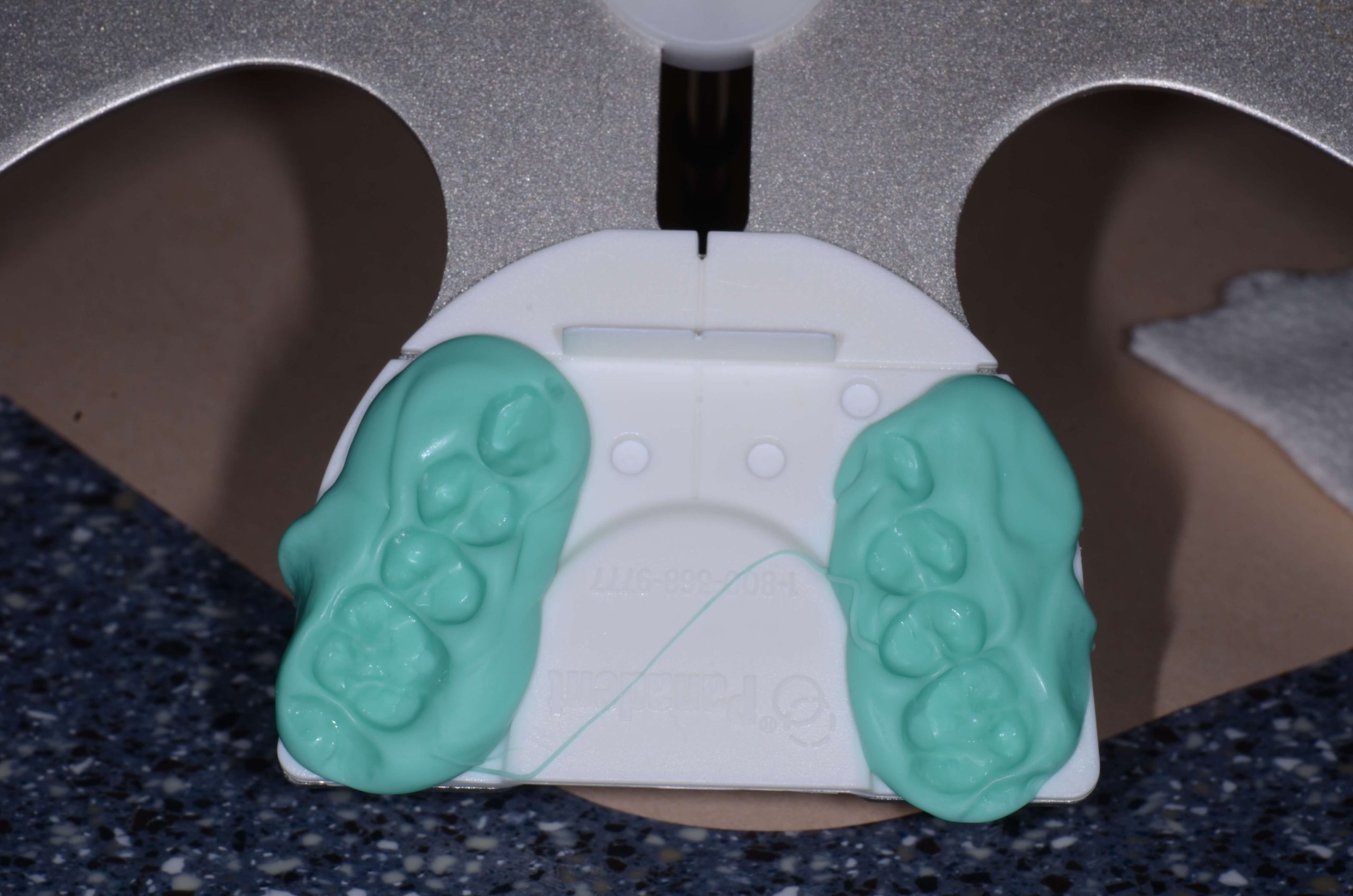
Once you have the white disposable plate – which is actually the piece you will send to the lab once the record is captured – snapped onto the Dento-Facial Analyzer, use VPS tray adhesive to lightly coat the plastic tray. You are only going to do this from about the canine position posteriorly because you aren’t going to put silicone on the anterior portion of that bite plate.

Next, attach the vertical reference bar to the Dento-Facial Analyzer. Without bite registration on it, take it to the patient’s mouth and seat the central incisors exactly against the white plastic in the front labially.

Verify that you can hold this level to the horizon in two planes of space and that you can touch the patient’s teeth. If not, you might need to build up the posterior.

If you’ve verified this, put bite silicone on the plate from the canine position back, then seat it again, making sure the central incisors are seated labially against the white plastic …

I’ll round up this fun technique with Part 3 in the series coming soon.

For a hands-on lesson in the Dento-Facial Analyzer from our talented educators, check out our Essentials 1 Pankey course. Also, watch this video for a quick refresher or pre-course overview.  

Related Course

E2: Occlusal Appliances & Equilibration

DATE: May 3 2025 @ 8:00 am - May 7 2026 @ 2:30 pm

Location: The Pankey Institute

CE HOURS: 44

Dentist Tuition: $ 7500

Single Occupancy with Ensuite Private Bath (per night): $ 355

What if you had one tool that increased comprehensive case acceptance, managed patients with moderate to high functional risk, verified centric relation and treated signs and symptoms of TMD? Appliance…

Learn More>

About Author

User Image
Lee Ann Brady DMD

Dr. Lee Ann Brady is passionate about dentistry, her family and making a difference. She is a general dentist and owns a practice in Glendale, AZ limited to restorative dentistry. Lee’s passion for dental education began as a CE junkie herself, pursuing lots of advanced continuing education focused on Restorative and Occlusion. In 2005, she became a full time resident faculty member for The Pankey Institute, and was promoted to Clinical Director in 2006. Lee joined Spear Education as Executive VP of Education in the fall of 2008 to teach and coordinate the educational curriculum. In June of 2011, she left Spear Education, founded leeannbrady.com and joined the dental practice she now owns as an associate. Today, she teaches at dental meetings and study clubs both nationally and internationally, continues to write for dental journals and her website, sits on the editorial board of the Journal of Cosmetic Dentistry, Inside Dentistry and DentalTown Magazines and is the Director of Education for The Pankey Institute.

FIND A PANKEY DENTIST OR TECHNICIAN

I AM A
I AM INTERESTED IN

VIEW COURSE CALENDAR

Panadent Dento-Facial Analyzer Technique: Introduction

September 21, 2018 Lee Ann Brady DMD

The Dento-Facial Analyzer is a marvelous tool I use in the practice to mount maxillary models. It has made a huge difference in my practice of dentistry and is one of my favorite tools to teach.

Introduction to the Dento-Facial Analyzer

For the critical aspects of diagnostics and sending info to the lab for the completion of a restorative case, mounting models appropriately is so important. They must be mounted in three planes of space referenced to hinge access to capture esthetic information including incisal plane and occlusal plane relative to the horizon.

Traditionally, this has been accomplished by utilizing a Facebow, Earbow, or by actually capturing hinge access position. Now, we have the option of using the Kois Dento-Facial Analyzer to capture both functional information and esthetic information that we would normally get with a Fox’s bite plane or stick bite. All of this functionality is managed with one simple device.

The Kois Dento-Facial Analyzer was designed based on scientific information gathered by Dr. John Kois, which shows that the distance from the incisal edge position of the maxillary central incisors to hinge access on average is 100 mm. Most people fall within a range of 5 mm to the average, therefore this is the assumption made when the device takes a record.

The armamentarium for record capturing with the Panadent instrument includes the analyzer, bite registration silicone in a gun with a tip, VPS adhesive used in an impression tray, and disposable bite plates that snap onto the analyzer (from the device manufacturer Panadent).

You can use bite registration silicone, Panadent bite tabs, wax, or VPS heavy body impression material to capture the record …

I’ll continue this review of the Dento-Facial Analyzer technique in Part 2, coming soon! And don’t miss one of my recent Pankey Gram favorites from Dr. Bill Gregg on an occlusion-focused hygiene exam. Read it here for his insightful tips.

For an in-person, hands-on lesson in the Dento-Facial Analyzer, check out our Essentials 1 Pankey course. You can also watch this video for a quick refresher.  

Related Course

Mastering Advanced Splint Therapy

DATE: November 13 2025 @ 8:00 am - November 16 2025 @ 1:00 pm

Location: The Pankey Institute

CE HOURS: 29

Dentist Tuition : $ 5900

Single Occupancy with Ensuite Private Bath (per night): $ 345

If you are ready to take what you know about appliance therapy to the next level, then this course is a must. The anatomic appliance is one of the most…

Learn More>

About Author

User Image
Lee Ann Brady DMD

Dr. Lee Ann Brady is passionate about dentistry, her family and making a difference. She is a general dentist and owns a practice in Glendale, AZ limited to restorative dentistry. Lee’s passion for dental education began as a CE junkie herself, pursuing lots of advanced continuing education focused on Restorative and Occlusion. In 2005, she became a full time resident faculty member for The Pankey Institute, and was promoted to Clinical Director in 2006. Lee joined Spear Education as Executive VP of Education in the fall of 2008 to teach and coordinate the educational curriculum. In June of 2011, she left Spear Education, founded leeannbrady.com and joined the dental practice she now owns as an associate. Today, she teaches at dental meetings and study clubs both nationally and internationally, continues to write for dental journals and her website, sits on the editorial board of the Journal of Cosmetic Dentistry, Inside Dentistry and DentalTown Magazines and is the Director of Education for The Pankey Institute.

FIND A PANKEY DENTIST OR TECHNICIAN

I AM A
I AM INTERESTED IN

VIEW COURSE CALENDAR

QuickSplint for Diagnostics and Restorative Applications

August 29, 2018 Pankey Gram

QuickSplint has many functions in general practice, but it is also a great resource overall in both diagnostics and restorative dentistry. It’s ideal for patients who grind, those with endodontic issues, and anyone needing a restoration.

QuickSplint to Improve the Patient Experience

Many dentists see patients who have wear on their teeth but don’t think they grind at all. They might have said that they used to grind their teeth, but they believe they don’t anymore. You can use the QuickSplint as a learning experience where the patient is able to recognize the cause of the attrition.

It’s nice to rely on this quick, easy, and inexpensive device. Go ahead and have the patient sleep in it for a little while. The QuickSplint will then reveal whether or not they are clenching and grinding. After the proof is clearly demonstrated, you can have an impactful conversation with your patient.

Another option for QuickSplint use is as a means for handling parafunction and restorative materials selection. Today, we talk a lot about posterior materials when we are doing crowns on first or second molars. Are we going to use high strength ceramics or traditional PFM restorations? Instead of guessing about the patient’s parafunctional risk and how much load they will place on the restoration, you can easily get that information with a QuickSplint.

Additionally, you can use the QuickSplint as a post-op device after an endodontic procedure. You can reduce a patient’s discomfort significantly in less than three minutes and also help the tooth heal without occlusal pressure. You then won’t have to take the entire occlusal table off the problem tooth. This can even work during pre-op for a patient whose tooth appears to need endodontic treatment.

In our Essentials One course at Pankey we use the Quicksplint as an overnight deprogrammer to allow us to capture very accurate diagnostic records.

Do you use QuickSplint in your dental practice? Tell us your best tips for using this handy little tool!

Related Course

E2: Occlusal Appliances & Equilibration

DATE: March 27 2026 @ 8:00 am - March 31 2026 @ 2:30 pm

Location: The Pankey Institute

CE HOURS: 44

Dentist Tuition: $ 7500

Single Occupancy with Ensuite Private Bath (per night): $ 355

What if you had one tool that increased comprehensive case acceptance, managed patients with moderate to high functional risk, verified centric relation and treated signs and symptoms of TMD? Appliance…

Learn More>

About Author

User Image
Pankey Gram

FIND A PANKEY DENTIST OR TECHNICIAN

I AM A
I AM INTERESTED IN

VIEW COURSE CALENDAR