The Effect of Rubber Dam Isolation on Bond Strength to Enamel 

March 13, 2024 Christopher Mazzola, DDS

Christopher Mazzola, DDS 

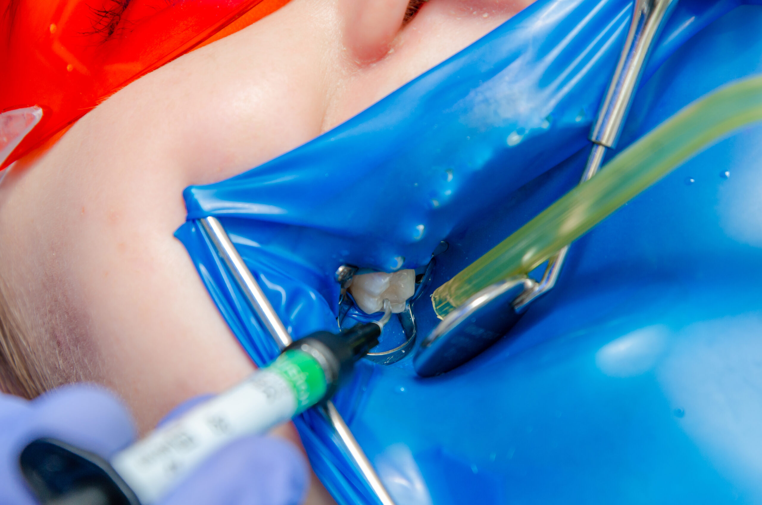
This is an example of a clinical study that can help us in our everyday practice of dentistry. Although the findings do not surprise us, keeping the findings in mind will guide us in decisions we make when performing treatments our patients are counting on to be long lasting. 

Dr. Markus Blatz is co-founder and past President of the International Academy for Adhesive Dentistry (IAAD) and Chairman of the Department of Preventive and Restorative Sciences and Assistant Dean for Digital Innovation and Professional Development at the University of Pennsylvania School of Dental Medicine in Philadelphia. He and a research team from the University of Coimbra, in Portugal, studied the effect of rubber dam isolation on bond strength to enamel. Their goal was to test two hypotheses. 

Hypothesis 1: Rubber dam isolation improves sheer bond strength independent of the adhesive system used. 

Hypothesis 2: A highly filled 3-step etch and rinse adhesive will provide higher bond strength values than an isopropyl-based universal adhesive. 

For their tests, they used OptiBond FL from Kerr for the 3-step etch and rinse adhesive and Prime & Bond Universal Adhesive for the isopropyl-based universal adhesive. 

The mesial, distal, lingual, and vestibular enamel surfaces of thirty human third molars were prepared (total n = 120 surfaces). A custom splint was made to fit a volunteer’s maxilla, holding the specimens in place in the oral cavity. Four composite resin cylinders were bonded to each tooth with one of two bonding agents (OptiBond FL and Prime & Bond) with or without rubber dam isolation. Shear bond strength was tested in a universal testing machine and failure modes were assessed. 

Both hypotheses were supported by the results reported in the Journal of Esthetic and Restorative Dentistry in November of 2022. 

  • With the rubber dam in place, both of the adhesives performed better than without the rubber dam in place, resulting in approximately twice as much shear bond strength with the rubber dam. 
  • The 3-step OptiBond FL system resulted in a more resilient bond than the Prime & Bond Universal adhesive. The OptiBond FL group with rubber dam presented the highest mean bond strength values. Fracture modes for specimens bonded without rubber dam isolation were adhesive and cohesive within enamel, while rubber dam experimental groups revealed only cohesive fractures. 

For the benefit of our patients, we shouldn’t cut corners that will impact the longevity of a restoration. My thoughts are that whenever we have basic pure enamel bonding it should be under a rubber dam, using a total etch, 3-step adhesive system. But considering dentin likes to be moist, we may need to make other clinical judgments.  

Related Course

Mastering Dental Photography: From Start to Finish

DATE: October 29 2026 @ 8:00 am - October 31 2026 @ 12:00 pm

Location: The Pankey Institute

CE HOURS: 19

Regular Tuition: $ 2995

Single Occupancy with Ensuite Private Bath (per night): $ 355

Dental photography is an indispensable tool for a high level practice. We will review camera set-up and what settings to use for each photo. All photos from diagnostic series, portraits,…

Learn More>
About Author

Default Avatar
Christopher Mazzola, DDS

FIND A PANKEY DENTIST OR TECHNICIAN

I AM A
I AM INTERESTED IN

VIEW COURSE CALENDAR

How Ivoclean Works 

March 1, 2024 Lee Ann Brady

How Ivoclean Works 

Lee Ann Brady, DMD 

Saliva on the inside of restoration surfaces greatly reduces the bond strength between the porcelain and the cement but during the intraoral try-in process, it is inevitable that there will be saliva contamination. 

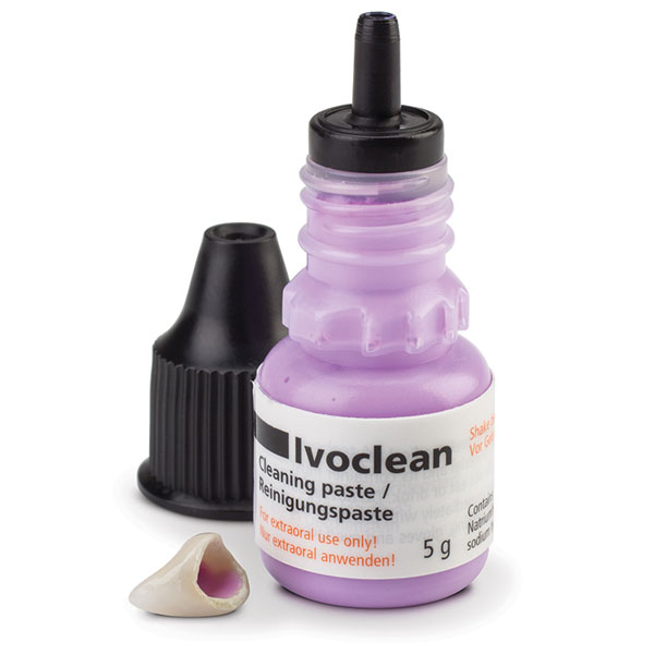
Most dentists I know use Ivoclean from Ivoclar to clean their indirect restorations after try-in. It is an incredible material for removing saliva and other contaminants that the restoration is exposed to during the intraoral try-in process.  

We trust Ivoclean to fully remove resin or traditional cements, as well as saliva and red blood cells to produce a super pristine surface.  

Did you ever wonder how Ivoclean works to get rid of saliva and all the other debris that gets on the inside of a ceramic restoration or metal base?  

Intraoral contaminants contain lots of phosphates. Ivoclean contains suspended zirconia particles that have an affinity for phosphates. The zirconia particles pull towards them the phosphate-laden particles, so when you rinse off the Ivoclean, the intraoral debris is rinsed away leaving a clean surface. 

Note: We don’t want to expose zirconia restorations to something that contains phosphates or includes phosphoric on the label because there is a strong attraction at an elemental level between zirconia and phosphate particles. To neutralize the ionic bond between saliva phosphates and zirconia, we need an alkaline solution such as potassium hydroxide (KOH). This is the active ingredient in products such as ZirClean from BISCO. 

Related Course

Mastering Dental Photography: From Start to Finish

DATE: October 29 2026 @ 8:00 am - October 31 2026 @ 12:00 pm

Location: The Pankey Institute

CE HOURS: 19

Regular Tuition: $ 2995

Single Occupancy with Ensuite Private Bath (per night): $ 355

Dental photography is an indispensable tool for a high level practice. We will review camera set-up and what settings to use for each photo. All photos from diagnostic series, portraits,…

Learn More>

About Author

User Image
Lee Ann Brady

FIND A PANKEY DENTIST OR TECHNICIAN

I AM A
I AM INTERESTED IN

VIEW COURSE CALENDAR

Why Use an Essix Retainer Versus a Flipper During Dental Implant Therapy

February 16, 2024 Lee Ann Brady DMD

Why Use an Essix Retainer Versus a Flipper During Dental Implant Therapy 

Lee Ann Brady, DMD 

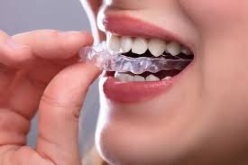
When it comes to choosing a provisional during implant therapy in the anterior aesthetic zone, we have two removable options. One is called a “flipper.” It’s an interim partial denture composed of an acrylic base and a denture tooth. The other is an Essix retainer.  

There is no question that both options are taxing for the patient for the three to five months that the patient is edentulous and must deal with having this removable device to replace the tooth. So, I always tell my patients that they are going to have to manage the provisional for that time, but it’s worth it because, in the end, they have replaced the tooth with an implant with all the benefits of an implant versus an alternative prosthetic solution. 

In my practice, I use Essix retainers in nearly 100% of the cases. Why? Because an Essix retainer is tooth-borne. The pressure is placed on the teeth and not on the surgical site. In the case of a flipper, the prosthesis is primarily tissue-borne with a little pressure placed on the adjacent teeth. We really don’t want any pressure on the surgical site while it is healing. Pressure can induce biological problems in bone grafts and connective tissue, which affect the long-term outcome. From an aesthetic perspective, the most challenging thing about anterior implant aesthetics is replicating the size, shape, and position of the tissues of the alveolar ridge and papilla. I want to do everything I can to eliminate pressure on the healing tissue. 

In my practice, we do Essix retainers that don’t have a full solid tooth in them. Instead, we simply paint flowable on the facial so that there’s zero pressure anywhere around that surgical site after extraction, after grafting, and after implant placement.  

In addition to explaining the improved outcomes associated with using an Essix retainer, I assure my patients that the retainer will be more comfortable to wear than a denture and be easily removed by them for eating, for drinking liquids other than water that are likely to stain the retainer, for teeth cleaning, and for cleaning the prosthesis. When out in public, such as in a restaurant, patients may carefully eat while wearing the Essix retainer.  

Related Course

Mastering Dental Photography: From Start to Finish

DATE: October 29 2026 @ 8:00 am - October 31 2026 @ 12:00 pm

Location: The Pankey Institute

CE HOURS: 19

Regular Tuition: $ 2995

Single Occupancy with Ensuite Private Bath (per night): $ 355

Dental photography is an indispensable tool for a high level practice. We will review camera set-up and what settings to use for each photo. All photos from diagnostic series, portraits,…

Learn More>

About Author

User Image
Lee Ann Brady DMD

Dr. Lee Ann Brady is passionate about dentistry, her family and making a difference. She is a general dentist and owns a practice in Glendale, AZ limited to restorative dentistry. Lee’s passion for dental education began as a CE junkie herself, pursuing lots of advanced continuing education focused on Restorative and Occlusion. In 2005, she became a full time resident faculty member for The Pankey Institute, and was promoted to Clinical Director in 2006. Lee joined Spear Education as Executive VP of Education in the fall of 2008 to teach and coordinate the educational curriculum. In June of 2011, she left Spear Education, founded leeannbrady.com and joined the dental practice she now owns as an associate. Today, she teaches at dental meetings and study clubs both nationally and internationally, continues to write for dental journals and her website, sits on the editorial board of the Journal of Cosmetic Dentistry, Inside Dentistry and DentalTown Magazines and is the Director of Education for The Pankey Institute.

FIND A PANKEY DENTIST OR TECHNICIAN

I AM A
I AM INTERESTED IN

VIEW COURSE CALENDAR

The Transition to Digital Dentistry Part 2

January 17, 2024 John Cranham, DDS

When my daughter Kaitlyn (currently in through E2 at Pankey) finished dental school in 2020, I strongly recommended that she learn analog first, then once mastered, make the transition to digital. This lasted about four months. I learned rapidly that this generation sees things in the digital realm far better than we do. She reminded me that “she grew up with a screen in her hand.” 

We began to focus on her learning the concepts of occlusion, esthetics, biology, tooth-by-tooth structural integrity, and visualizing and planning in the virtual (digital world). We quickly learned that, although she could easily visualize things on the computer, the patient is ultimately analog. We began to utilize an analog articulator for her to learn the hand skills of what we would do on the patient. 

A great example of this is equilibration. A “trial equilibration” on a virtual articulator is a 5-minute process that lets us determine if equilibration is an appropriate treatment option. The problem is that, unlike analog, you do not learn the brush strokes that will be required to perform this skill in the mouth. I have performed hundreds if not thousands of equilibrations. I know the brush strokes. For me, once I see on the virtual articulator that I can do the equilibration without too much tooth structure removal, I am ready to go to the mouth. For Kaitlyn, who has very limited equilibration experience, once visualized on the virtual articulator, then it’s time to go back to analog. She mounts the printed models on an analog articulator to perform a traditional trial equilibration. In this way, she learns the brushstrokes of this incredibly important procedure. 

I think it is extremely important that dentists, who are learning to equilibrate intraorally, work on mounted analog models to develop their equilibration skills. 

Returning to the consideration of the financial cost of bringing new technology into your practice—input devices (scanners and CBCTs), output devices (printers and mills), and software to manipulate the data all cost money. Doctors that are going down this road usually like technology and consider the dramatic increases in efficiency to ultimately increase the productivity and profitability of the practice. This is certainly something I have seen. The bottom line is dental stone will go away. We all must make the decision when it is appropriate to make the jump. 

Dr. Lee Ann Brady has invited me to audit all the Pankey Essentials courses over the next year. I am super excited about this. She has asked me to recommend ways to appropriately implement examples of digital technologies and workflows into these core classes. While younger dentists are drawn to digital information, it is important for us to remind them that our patients are ANALOG. We are training dentists to perform complex procedures on patients, not on computers. This requires great study and a commitment to understand timeless concepts, while simultaneously developing the hand skills to accomplish these procedures accurately and use digital workflows to make things more efficient. 

In 2024, The Pankey Institute is also implementing a digital hands-on course for those doctors who would like to make the transition over to virtual articulation and digital workflows—something that I am excited to be part of. Dentistry is in a great transition. I look forward to making sure the concepts that we have all built our practices around do not get lost in the digital world. 

About Author

User Image
John Cranham, DDS

Dr. John Cranham practices in Chesapeake, Virginia focusing on esthetic dentistry, implant dentistry, occlusal reconstruction, TMJ/Facial Pain and solving complex problems with an interdisciplinary focus. He practices with his daughter Kaitlyn, who finished dental school in 2020. He is an honors graduate of The Medical College of Virginia in 1988. He served the school as a part time clinical instructor from 1991-1998 earning the student given part time faculty of the year twice during his stint at the university. After studying form the greats in occlusion (Pete Dawson & The Pankey Institute) and Cosmetic Dentistry (Nash, Dickerson, Hornbrook, Rosental, Spear, Kois) during the 1990’s, Dr. Cranham created a lecture in 1997 called The Cosmetic Occlusal Connection. This one day lecture kept him very busy presenting his workflows on these seemingly diametrically opposed ideas. In 2001 he created Cranham Dental Seminars which provided, both lecture, and intensive hands on opportunities to learn. In 2004 he began lecturing at the The Dawson Academy with his mentor Pete Dawson, which led to the merging of Cranham Dental Seminars with The Dawson Academy in 2007. He became a 1/3 partner and its acting Clinical Director and that held that position until September of 2020. His responsibilities included the standardization of the content & faculty within The Academy, teaching the Lecture Classes all over the world, overseeing the core curriculum, as well as constantly evolving the curriculum to stay up to pace with the ever evolving world of Dentistry. During his 25 years as an educator, he became one of the most sought after speakers in dentistry. To date he has presented over 1650 full days of continuing education all over the world. Today he has partnered with Lee Culp CDT, and their focus is on integrating sound occlusal, esthetic, and sound restorative principles into efficient digital workflows, and ultimately coaching doctors on how to integrate them into their practices. He does this under the new umbrella Cranham Culp Digital Dental. Dr. Cranham has published numerous articles on restorative dentistry and in 2018 released a book The Complete Dentist he co-authored with Pete Dawson. In 2011 He along with Dr. Drew Cobb created The Dawson Diagnostic Wizard treatment planning software that today it is known as the Smile Wizard. Additionally, He has served as a key opinion leader and on advisory boards with numerous dental companies. In 2020 he published a book entitled “The Cornell Effect-A Families Journey Toward Happiness, Fulfillment and Peace”. It is an up from the ashes story about his adopted son, who overcame incredible odds, and ultimately inspired the entire family to be better. In November of 2021 it climbed to #5 on the Amazon best seller list in its category. Of all the things he has done, he believes getting this story down on paper is having the greatest impact.

FIND A PANKEY DENTIST OR TECHNICIAN

I AM A
I AM INTERESTED IN

VIEW COURSE CALENDAR

The Transition to Digital Dentistry – Part 1 

October 18, 2023 John Cranham, DDS

When dentists are considering the purchase of a new digital technology for implementation in their clinical practice, they usually have three primary considerations:

  • Will it increase accuracy?
  • Will it increase consistency?
  • Is it possible to increase efficiency without sacrificing quality?

Often the cost is a major consideration, and I will come back to the question of cost later in this two-part series.

Twenty years ago, with first-generation CEREC, the results were not close to what we could do in the analog world. But I jumped into using chairside CAD/CAM because, at the time, I was teaching with Dr. Peter Dawson and many doctors were buying the technology. We needed to be able to talk about how it fit with complete-care dentistry.

It was a difficult time because while we could do crowns (usually one or two at a time) in maximum intercuspation or on equilibrated patients in CR, the software was not sophisticated enough to do any kind of case planning. It did, however, provide me with the first glimpse at scanning and 3D digital technology, and I became excited about the possibilities. I started to think about the ability to scan mouths for diagnostic purposes and to do waxups. Virtual articulation was not there, and we didn’t have 3D printing to print our “outputs” so I continued to wait.

As recently as four years ago, my son would spend a day mounting stone models for me. Today, he has digital models mounted on a virtual articulator before the patient leaves their appointment. Digital and AI software platforms are evolving quickly. They enable more dentists and lab technicians to visualize optimal dentistry and design occlusions and beautiful smiles easily. For implant dentistry and orthodontics, these digital and AI software platforms remove obstacles and inaccuracies.

The ability to “stack” data sets on top of one another (pre-op model, waxup, CBCT, Face Scans, photos) allows us to see things in ways I could have never imagined. The things I dreamed about 15 years ago are here now and evolving at warp speed. I honestly don’t remember a time when I was more excited about day-to-day dentistry. A special shout out to Lee Culp (Sculpture Studios), who introduced me to these workflows, and continues to lead dentistry into the digital era.

In the digital world, we can make digital impressions, face-bows, and waxups to see if we need to equilibrate, orthodontically move teeth, or change the shape/contour of the teeth. Technology allows us an efficient, accurate workflow. An experienced digital team can mount models and a virtual articulator, in CR, in 10-15 minutes. The ability to scan in CR, MI, or any treatment position can be accomplished with ease. Trial equilibrations or diagnostic designs (waxups) also can be accomplished far more rapidly, with beautiful results.

As digital workflows take hold in mainstream dentistry, one of the great challenges we face is how the next generations of dentists will learn. In treatment planning, I have the benefit of having 30-plus years of experience working in analog. I have solidified the concepts of centric relation, anterior guidance, posterior disclusion, crossover, envelope of function, incisal edge position, and all the things that go along with visualizing optimum care for your patent. This knowledge was my foundation for determining if these technologies had evolved enough to make the shift to the digital realm.

About Author

User Image
John Cranham, DDS

Dr. John Cranham practices in Chesapeake, Virginia focusing on esthetic dentistry, implant dentistry, occlusal reconstruction, TMJ/Facial Pain and solving complex problems with an interdisciplinary focus. He practices with his daughter Kaitlyn, who finished dental school in 2020. He is an honors graduate of The Medical College of Virginia in 1988. He served the school as a part time clinical instructor from 1991-1998 earning the student given part time faculty of the year twice during his stint at the university. After studying form the greats in occlusion (Pete Dawson & The Pankey Institute) and Cosmetic Dentistry (Nash, Dickerson, Hornbrook, Rosental, Spear, Kois) during the 1990’s, Dr. Cranham created a lecture in 1997 called The Cosmetic Occlusal Connection. This one day lecture kept him very busy presenting his workflows on these seemingly diametrically opposed ideas. In 2001 he created Cranham Dental Seminars which provided, both lecture, and intensive hands on opportunities to learn. In 2004 he began lecturing at the The Dawson Academy with his mentor Pete Dawson, which led to the merging of Cranham Dental Seminars with The Dawson Academy in 2007. He became a 1/3 partner and its acting Clinical Director and that held that position until September of 2020. His responsibilities included the standardization of the content & faculty within The Academy, teaching the Lecture Classes all over the world, overseeing the core curriculum, as well as constantly evolving the curriculum to stay up to pace with the ever evolving world of Dentistry. During his 25 years as an educator, he became one of the most sought after speakers in dentistry. To date he has presented over 1650 full days of continuing education all over the world. Today he has partnered with Lee Culp CDT, and their focus is on integrating sound occlusal, esthetic, and sound restorative principles into efficient digital workflows, and ultimately coaching doctors on how to integrate them into their practices. He does this under the new umbrella Cranham Culp Digital Dental. Dr. Cranham has published numerous articles on restorative dentistry and in 2018 released a book The Complete Dentist he co-authored with Pete Dawson. In 2011 He along with Dr. Drew Cobb created The Dawson Diagnostic Wizard treatment planning software that today it is known as the Smile Wizard. Additionally, He has served as a key opinion leader and on advisory boards with numerous dental companies. In 2020 he published a book entitled “The Cornell Effect-A Families Journey Toward Happiness, Fulfillment and Peace”. It is an up from the ashes story about his adopted son, who overcame incredible odds, and ultimately inspired the entire family to be better. In November of 2021 it climbed to #5 on the Amazon best seller list in its category. Of all the things he has done, he believes getting this story down on paper is having the greatest impact.

FIND A PANKEY DENTIST OR TECHNICIAN

I AM A
I AM INTERESTED IN

VIEW COURSE CALENDAR

Why Use a Dental Microscope? 

August 14, 2023 Michelle Lee DDS

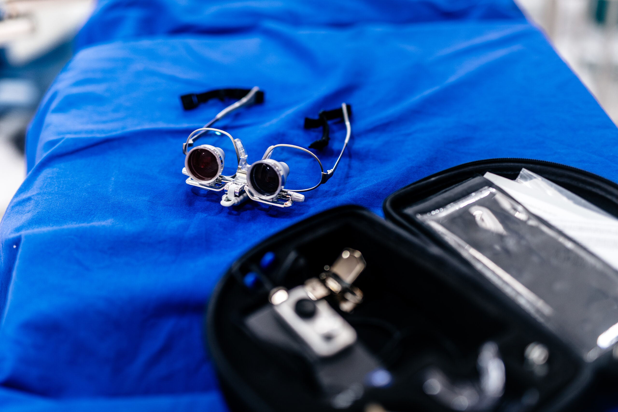
We have a buffet of restorative materials to choose from today. Accurate preparations are fulfilling the promise of digital design and precision-based dentistry to deliver the highest level of restorative treatment with efficiency. The microscope has changed the way I personally approach dentistry, and it has heightened my passion for precision dentistry.

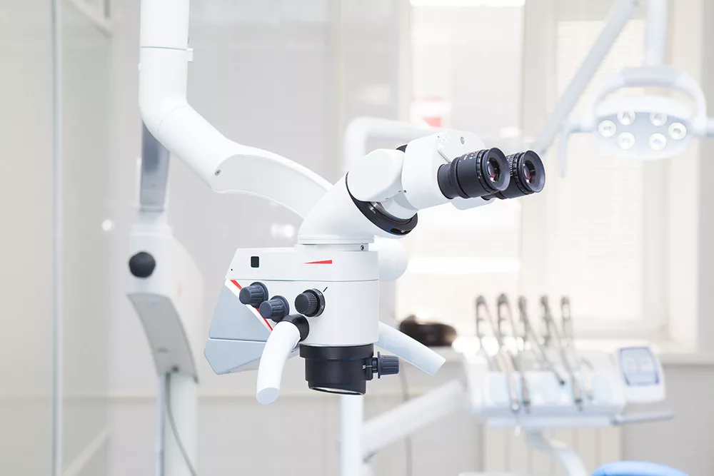
The world of dental microscopy has opened my eyes to see and know more. The microscope has allowed me to improve my dentistry through ergonomics, improved illumination, and magnification. I am astonished and, at the same time, feel humbled to be able to see detail and precision at such a high level of magnification.

An Opportunity to Teach Propelled Me

My introduction to using a dental microscope began five years ago through the University of Pennsylvania Dental School where I was invited to teach a novel program introducing the idea of restorative dentistry with the utilization of dental microscopes. The purpose of the program was to introduce concepts of precision-based dentistry in our ever-changing digital dental world. We wanted our students to appreciate the value of details and how marginal gaps and adaptation can be improved with a higher level of thinking and training.

Not owning a microscope at the time, I agreed to explore this idea and accepted the position. I trained, calibrated, and practiced with the dental students to quickly see the value. Within a few short months, I had integrated dental microscopy into my dental practice.

Does the Microscope Replace Loupes?

Other dentists, who have not adopted dental microscopy, often ask me, “What does the microscope replace in your practice, and does it replace your loupes?” The answer is simple. Using a dental microscope doesn’t necessarily replace loupes but higher magnification increases proficiency and precision. I often prepare with my loupes and finish my preparations under higher magnification to improve smoothness, adaptation, and finish lines.

The viewing capabilities of the microscopes provide a range of higher magnification beyond some loupes with no eye muscle strain. Under dental microscopy, I can magnify my viewing field from 4X power to 10X power and sometimes as high as 25X power.

The Microscope Has Changed How I Practice

Utilizing a dental microscope has changed how I practice dentistry, improved my overall health in ergonomics, and is now an invaluable part of my practice as I strive to serve my patients with higher-level dentistry.

Using the microscope, I routinely minimize marginal gaps in my preparations to increase the longevity of restorations for my patients.

The completely upright binocular, parallel vision provides less strain to my posture, my neck, and head position.

My patients can view what I see with the microscope and gain an elevated understanding. Together we can partner better in making collaborative decisions to improve their dental health in the best way possible.

Why I Use a Dental Microscope

I am of the belief that when we see better, we can do better. I want to do my best for my patients. The dental microscope provides an elevated level of magnification, illumination, ergonomics, and patient education so I can deliver the highest care. After adapting to its usage and experiencing its benefits, I recommend it highly to other dentists. If you have access to one to try it–and put in the effort to learn how to use it, I think you’ll rapidly want one of your own.


Discover more on how to build a thriving dental practice with The Essentials Series at Pankey. This comprehensive 4-part course starts with Essentials 1, diving deep into the core principles that will transform your approach to patient care and practice management. 

Related Course

E1: Aesthetic & Functional Treatment Planning

DATE: July 9 2026 @ 8:00 am - July 12 2024 @ 2:30 pm

Location: The Pankey Institute

CE HOURS: 39

Dentist Tuition: $ 6900

Single Occupancy with Ensuite Private Bath (Per Night): $ 355

Transform your experience of practicing dentistry, increase predictability, profitability and fulfillment. The Essentials Series is the Key, and Aesthetic and Functional Treatment Planning is where your journey begins.  Following a system of…

Learn More>

About Author

User Image
Michelle Lee DDS

Dr. Michelle Lee is very proud to provide all aspects of general, family, and cosmetic dentistry to the Fleetwood and Berks county areas. Dr. Lee is a 2004 graduate of the University of Pennsylvania School of Dental Medicine and completed a one-year General Practice Residency program at Abington Memorial Hospital. Dr. Lee continues to keep herself abreast of dental advancements and takes hundreds and hundreds of hours of advanced dental education from the Pankey Institute and other courses for advanced dental training. She also maintains a faculty and advisor position at the Pankey Institute. Professionally, Dr. Lee is member of the Academy of General Dentistry, American Dental Association, Pennsylvania Dental Association, and serves on a committee of the American Equilibration Society. She also volunteers to treat pediatric patients through her local dental society.

FIND A PANKEY DENTIST OR TECHNICIAN

I AM A
I AM INTERESTED IN

VIEW COURSE CALENDAR

Personalized Precision Implant Dentistry

July 26, 2023 Michael J. Costa, DDS, MHA

It’s not “uncommon” for a private practice that is dedicated to personalized, comprehensive care with a mostly fee-for-service patient base to develop one or more “niche” services that distinguish it from other practices. For example, they might develop expertise in Invisalign, Airway Management, Integrated Dental Medicine, Microscopic Dentistry, or in my case, Personalized Implant Dentistry.

Passion and Purpose

I have a passion for what I do, which is making a difference in people’s lives with implant treatment and the care and attention I give them. Each patient is special to me, and I want to solve their dental problems. Becoming an associate in the practice of a Pankey Scholar and faculty member, Dr. Steven Malone, has been an incredible experience. Our relationship has been synergistic from the start! He has mentored and supported me as I built an implant practice inside his comprehensive general dentistry practice and became known in the Knoxville area as a person-centered implant dentist. With the implant practice and a full-service in-house dental lab inside our Knoxville Smiles location, Steve and I offer more comprehensive dental services under one roof than most private dentists.

Invested in Best Care

At Knoxville Smiles, we have invested in state-of-the-art equipment because it enables us to provide better, safer care and improved outcomes, but that is not the only thing that sets us apart. We genuinely treat our patients as members of our family and provide compassionate, personalized care in a comfortable environment designed to make them feel safe and at home. We take things as slowly or as quickly as each patient desires. We spend as much time with each patient as they need. And, because we are known for this, we have no shortage of patients who place priority on health and collaboratively participate in diagnosis and treatment planning. We rarely have a patient who declines to move forward or stalls out with achieving the dentistry they need and a naturally beautiful smile.

Recently, we invested in a robotically-guided dental implant surgery assistant called “Yomi.” It uses haptic guidance and comprehensive information about the patient’s oral and facial structures to guide the precise placement of implants. What “sold” me on the investment was that it provides real-time, multi-sensory guidance. With it, I can monitor nerve locations and other orofacial structures, so I can immediately alter treatment based on real-time information. With this greater precision, the implant surgical experience is less-invasive.

My experience, in the couple months that I have been using Yomi, is that the implant placement procedure takes less time, and the desired results are more predictable. We have made a point in our implant marketing to mention our “latest technology” and new patients who call say they like the idea of having their dental implant treatment done with Yomi.

Much of the Fun Is in the Adventure

If you are a dentist working in a person-centered general practice who has the desire to develop a niche within the practice, my advice is to go ahead and develop the knowledge and expertise it will take. The road to get you to where you want to go will be eventful and personally rewarding. Much of my implant knowledge and experience was gained while in the military, I completed the Augusta University Comprehensive Training in Implant Dentistry Course, and I am currently working on becoming an Associate Fellow of the American Academy of Implant Dentistry. I believe I will always be on the road to mastery because it is so meaningful. My “personalized” approach to clinical patient care is constantly being honed as I progress through The Pankey Institute curriculum and am mentored by Dr. Steven Malone. Can you tell I love what I do?

See you at The Pankey Symposium!

Related Course

E3: Restorative Integration of Form & Function

DATE: November 1 2026 @ 8:00 am - November 5 2026 @ 2:30 pm

Location: The Pankey Institute

CE HOURS: 41

Dentist Tuition: $ 7500

Single Occupancy with Ensuite Private Bath (per night): $ 355

Understanding that “form follows function” is critical for knowing how to blend what looks good with what predictably functions well. E3 is the phase of your Essentials journey in which…

Learn More>

About Author

User Image
Michael J. Costa, DDS, MHA

Dr. Michael Costa practices in Knoxville, TN with his business partner Dr. Stephen Malone. Dr. Costa graduated from the UNC Adams School of Dentistry in 2014 after completing his Masters in Healthcare Administration in 2010. After graduation he completed his AEGD in the US Navy where he served active duty for 3 years. Dr. Costa started using the Yomi Robot in 2021 and quickly became a Key Opinion Leader. Dr. Costa is a Pankey Alumni. Dr. Costa resides in Knoxville with his wife, Dr. Bre and their two children Bennett and Bella.

FIND A PANKEY DENTIST OR TECHNICIAN

I AM A
I AM INTERESTED IN

VIEW COURSE CALENDAR

Using Digital Technology to Create an Analog Smile Mock-Up with Your Patient

February 21, 2022 Daren Becker DMD

Today we move between the digital and the analog world to accomplish the goals of aesthetic dentistry. A mock-up is a key tool in helping patients want aesthetic dentistry and visualizing what the changes will accomplish.

Lots of us have learned from masters like Dr. Susan Hollar how to hand-lay composite on the patient’s teeth so the patient can see their possible new smile. This trial smile technique is a fabulous way to motivate patients. It’s also a great way for us to learn what might be possible.

For many dentists, that technique is not natural for us, and it takes chair time. Another way we can model possible changes is through digital technology. In our office, we are using digital smile design as follows.

1. We do our initial records, which includes facial photos and an intraoral scan using our digital impressions intraoral scanning system.

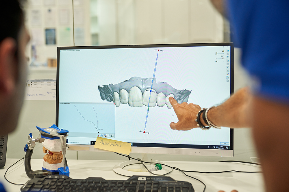
2. Either on the software in our office or at the lab, a 3-D version can be designed of what the new smile approximately could look like.This doesn’t have to be a definitive wax-up. Remember, we call it a diagnostic work-up. In fact, this is oftentimes where we discover the need for gingival changes and/or orthodontic procedures in order to achieve the desired outcome. I find this extremely helpful in communicating with the patient as I can show them what the compromised outcome would be if they choose not to correct the gingival levels or align the teeth if that is in fact appropriate.

We’ve learned it is very efficient to collaborate with the lab, the lab creates the 3-D design, and the lab emails us the STL digital file of the design. Alternatively, the lab can send printed models, matrices, or even milled/printed PMMA shells of the design.

3. On the 3-D printer in our office, we print the model from the STL file.

4. We make a matrix from that, either in a suck down material or a putty matrix, and we take that to the mouth, fill it with our temporary material (usually bisacryl), and seat it right onto the teeth.

5. After letting it set, removing the matrix, and peeling off excess material, the patient is wearing their trial smile. This last step takes all of two minutes.

Using this process enables us to do the lab work between appointments, and when the patient returns, they can very quickly preview the possibilities.

It is a wonderful communication tool, because the patient can look in their own mouth, not at a picture of someone else, not at pictures of other shapes of teeth, and say, “I like that,” or “I thought they would be shorter (longer, fatter, narrower…).” You can go in with your handpiece and reshape the temporary material or add material with flowable to make something more pronounced.

Patient participation in the tweaking of the design draws the patient into deeper engagement with and commitment to the smile they want. Now, we can scan the corrected and approved trial smile while it is in their mouth and take photos to send to the lab to help them as we move into the definitive design phase, including working out the occlusion and function.

Related Course

Functional Esthetic Excellence – Utilizing 100% Digital Workflow

DATE: May 8 2025 @ 8:00 am - May 10 2025 @ 2:00 pm

Location: The Pankey Institute

CE HOURS: 25

Dentist Tuition: $ 3195

Single Occupancy with Ensuite Private Bath (per night): $ 345

Embracing Digital Dentistry This course will introduce each participant to the possibilities of complex case planning utilizing 100% digital workflows. Special emphasis will be placed on understanding how software can…

Learn More>

About Author

User Image
Daren Becker DMD

Dr. Becker earned his Bachelors of Science Degree in Computer Science from American International College and Doctor of Dental Medicine from the University of Florida College of Dentistry. He practices full time in Atlanta, GA with an emphasis on comprehensive restorative, implant and aesthetic dentistry. Daren began his advanced studies at the Pankey Institute in 1998 and was invited to be a guest facilitator in 2006 and has been on the visiting faculty since 2009. In addition, in 2006 he began spending time facilitating dental students from Medical College of Georgia College of Dentistry at the Ben Massell Clinic (treating indigent patients) as an adjunct clinical faculty. In 2011 he was invited to be a part time faculty in the Graduate Prosthodontics Residency at the Center for Aesthetic and Implant Dentistry at Georgia Health Sciences University, now Georgia Regents University College of Dental Medicine (formerly Medical College of Georgia). Dr. Becker has been involved in organized dentistry and has chaired and/or served on numerous state and local committees. Currently he is a delegate to the Georgia Dental Association. He has lectured at the Academy of General Dentistry annual meeting, is a regular presenter at ITI study clubs as well as numerous other study clubs. He is a regular contributor at Red Sky Dental Seminars.

FIND A PANKEY DENTIST OR TECHNICIAN

I AM A
I AM INTERESTED IN

VIEW COURSE CALENDAR

Easy to Mount Printed Dental Models

February 14, 2022 Mark Kleive DDS

In my dental practice, we often use mounted models for diagnostics and restorative treatment planning. Three years ago, we began 3D printing these models.

One of the benefits of printed models is their higher durability compared to stone models. Also, long term, we do not need to save the printed models because we have the digital models saved in the patient’s file. But the greatest benefit has been the efficiency gained in mounting models on articulators. As a result, we have decreased our overhead and increased our mix of services.

What made this mounting efficiency possible is software called “Blue Sky Plan” from Blue Sky Bio. Blue Sky Plan is advanced dental treatment planning software used for milling and printing dental products. One of its applications is printing surgical implant guides, but it has many dental and medical applications for anatomical modeling, surface editing, and offsetting. It allows for CT scan importation and analysis, and export to STL format for 3D printing.

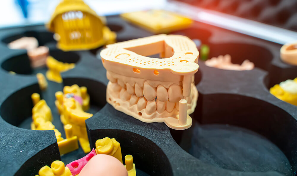
When dental models are printed, the interior can be hollow with a waffle pattern on the back that makes articulator mounting super easy. The process is as simple as opening the software, going to editing, importing your scan, and then selecting a hollow model with the waffle base. To print the waffle base on the model, you need to scan the entire pallet.

Related Course

E3: Restorative Integration of Form & Function

DATE: January 12 2025 @ 8:00 am - January 16 2025 @ 2:30 pm

Location: The Pankey Institute

CE HOURS: 41

Dentist Tuition: $ 7400

Single Occupancy with Ensuite Private Bath (per night): $ 345

THIS COURSE IS SOLD OUT Understanding that “form follows function” is critical for knowing how to blend what looks good with what predictably functions well. E3 is the phase of…

Learn More>

About Author

User Image
Mark Kleive DDS

Dr. Mark Kleive earned his D.D.S. degree with distinction from the University of Minnesota School of Dentistry in 1997. Mark has had experience as an associate in a multi-clinic setting and as an owner of 2 different fee-for-service practices. For the last 6 years Mark has practiced in a beautiful area of the country – Asheville, North Carolina, where he lives with his wife Nicki and twin daughters Meighan and Emily. Mark has been passionate about advanced education since graduation. Mark is a Visiting Faculty member with The Pankey Institute and a 2015 inductee into the American College of Dentistry. He leads numerous small group study clubs, lectures nationally and offers his own small group programs. During the last 19 years of practice, Dr. Kleive has made a reputation for himself as a caring, comprehensive oral healthcare provider.

FIND A PANKEY DENTIST OR TECHNICIAN

I AM A
I AM INTERESTED IN

VIEW COURSE CALENDAR

From Your Practice to the Lab – Continuation of a Philosophy of Treatment

May 28, 2021 Josh Polansky

This blog is a precursor for the long lecture I will do on this topic at the 2021 Pankey Symposium.

Over the last decade, there have been major changes in how we do things in our laboratory (Niche Dental Studio), but from small cases to full mouth and hybrid cases, traditional Prosthodontic protocols still guide everything we do. These foundational processes provide a structural/philosophical approach for all our cases.

It’s a philosophical approach to diagnosis and treatment that you have been learning in your Pankey Institute courses. It’s an approach that extends from your dental practice into our lab, so our lab becomes part of your practice.

What are the key principles of this philosophy or approach?

  • We will use optimal diagnostic protocols, communication, technology and methods to deliver custom prostheses as efficiently as possible while not compromising on the quality of the products.
  • We will do our best to deliver products that meet or exceed your expectations for optimal function, comfort, and natural esthetics.

Here are some of the things that we do the same and some we do differently than we did ten years ago.

Feldspathic ceramics still produce the most natural appearance.

In the past everything we made was made by hand, and it was the prosthodontic protocols of this handwork that enabled us to have success using CADCAM technology today. And while today’s CADCAM dentistry is great, it does not replicate the results of restorations made by hand. A machine can’t mill “infinity margins.” Monolithic materials used in milling do not contain multiple levels of opacity.

To blend perfectly with Nature, restorations must still be made by hand, and in our laboratory, feldspathic veneers are still our “go to” type of restoration for central incisors. Layered feldspathic ceramics not only look the best but also are the best for marginal integrity. The restoration on number 8 below is an example.


For fit and finish, these types of anterior restorations are still the prosthodontic foundation of our Niche Dental Studio.

We still aim to replicate natural teeth.

Another foundational attribute of prosthodontic protocols is to replicate nature. Part of our success has been how much time and effort we have put into studying natural teeth and helping Pankey Institute trained dentists distinguish themselves by using restorations that are exquisitely made to appear natural and blend in the patient’s smile.

Today’s patients desire natural esthetics once they understand the elements of what makes teeth appear natural. If a patient seems stuck on a cosmetic dentistry meme of the past and requests whiter, brighter, straight teeth that will not blend in their smile, a conversation with your patient that illustrates tooth, smile, and facial esthetics will be appreciated by your patient and distinguish you as a caring, exacting dentist.

To create restorations that appear natural and don’t “jump out,” we do the following things:

  • Increase the “value” of the color but not enough to create harsh contrast.
  • Play with the levels of the incisal embrasures and the translucency.

These prosthodontic protocols can be implemented by you, too, while doing composite build-ups.

We use new technology to optimize communication.

From the ceramist’s perspective, I don’t want to see just close-up images of teeth. I want to see the patient. For many of our cases, we see the patient in our lab. Local patients come in for a consultation. We consult with other patients via Skype or Facetime. Seeing the entire smile, the entire face in natural interaction, aids us in doing our best.

 Modern 3D technology has changed how labs communicate visually with doctors and their patients. We’re constantly sending 3D screen shots back and forth with our doctors so they can check out the design and show them to a patient. An image like this one is confusing to patients. So, we’ve been able to integrate those screen shots into a photo of the patient to create a virtual image the patient grasps more easily.

CAD technology allows us to work more efficiently, but we still hand-finish restorations.

In our laboratory, we mill a lot of lithium disilicate crowns for clients. Prior to milling the lithium disilicate, we like to mill the restorations in wax. The milling quickly does 80% of the model creation and gives us the opportunity to hand finish the other 20% as we traditionally would. We can now put all our esthetic and creative efforts into finishing the case. We also mill temporary restorations from IOS data without hand modifying them.

Using IOS and CAD has made the lives of our clients much easier. For example, in the past, with full mouth cases, we did a lot of wax-ups when raising verticals. The doctors found working with matrixes too time consuming. They preferred working with eggshells and would reline them. Little problems would creep in when seating these eggshells. Perhaps, the cant was a little off or the vertical wasn’t raised accurately. With 3D imaging, it is far easier, because now we can do our full mouth wax-ups, scan them, and print the eggshells from scans with full palatal rest and retro-molar rest. There is now only one definitive way to seat the eggshells.

This is just a taste. There is so much to share.

To see how we do actual cases, in detail, go to the free Pankey Webinar: Prosthodontic Protocols for the Modern Dental Team. There you will see how our modernized approach, guided by traditional prosthodontic processes, becomes an extension of your treatment goals. I look forward to sharing more with you at the 2021 Pankey Symposium.

Related Course

E3: Restorative Integration of Form & Function

DATE: February 8 2026 @ 8:00 am - February 12 2026 @ 2:30 pm

Location: The Pankey Institute

CE HOURS: 41

Dentist Tuition: $ 7500

Single Occupancy with Ensuite Private Bath (per night): $ 355

Understanding that “form follows function” is critical for knowing how to blend what looks good with what predictably functions well. E3 is the phase of your Essentials journey in which…

Learn More>

About Author

User Image
Josh Polansky

Joshua Polansky earned his Bachelor of Arts degree, Summa Cum Laude, from Rutgers University in 2004. While working part-time at a dental laboratory, he took advantage of an opportunity to apprentice with distinguished master technician, Olivier Tric of Oral Design Chicago. Mr. Tric opened Joshua’s eyes to a whole new world of possibilities. He made the decision to become a master dental technician following the path that Tric had forged. He continued to acquire technical skills by studying in Europe with other mentors and experts in the field such as Klaus Muterthies. Joshua earned his Masters degree in dental ceramics at the UCLA Center for Esthetic Dentistry under Dr. Edward McLaren. Joshua Continued his training under Jungo Endo and Hiroaki Okabe at UCLA’s advanced prosthodontics and maxillofacial program working on faculty and residents cases. Joshua currently resides in Cherry Hill, NJ where he is the owner and operator of Niche Dental Studio.

FIND A PANKEY DENTIST OR TECHNICIAN

I AM A
I AM INTERESTED IN

VIEW COURSE CALENDAR